
New Technology Helps Endocrine Surgeons Make Surgery Safer
Johns Hopkins surgeons are harnessing light-based technology to spot parathyroid tissue, protect nerve function and reduce surgical complications.
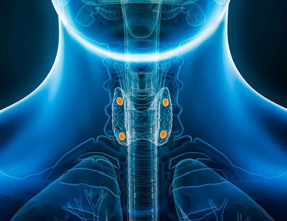
A new tool uses the natural autofluorescence of parathyroid glands (highlighted in orange) to help endocrine surgeons distinguish them from surrounding tissue during operations.
Johns Hopkins endocrine surgeons are advancing surgical care with innovative technology designed to make thyroid and parathyroid surgery safer, more precise and less invasive. One new tool helping them do that is the probe-based near-infrared autofluorescence (NIRAF) detection system. This tool uses the parathyroid glands’ natural autofluorescence to distinguish them from surrounding tissue. The Johns Hopkins Hospital is one of the only facilities in the state that has this technology.
The four parathyroid glands sit behind the thyroid and are each about the size of a grain of rice. The glands, which regulate calcium and phosphorus levels in the blood, can be hard to distinguish from fat and lymph nodes.
The NIRAF detection system serves as an adjunctive tool that can help during complex thyroid and parathyroid surgeries in which surgeons want to identify parathyroids in the face of challenging anatomy or preserve the parathyroid glands in cases such as thyroidectomies and reoperative surgeries for patients with thyroid cancer in which scar tissue in the neck makes the surgery more difficult.
“With this probe, we measure the background autofluorescence on thyroid tissue, and when we place it on parathyroid tissue, the signal is typically several orders of magnitude higher” says Lilah Morris-Wiseman, chief of the Johns Hopkins Division of Endocrine Surgery. “It gives us a fast and reliable way to confirm parathyroid tissue without injecting anything — just a quick, noninvasive readout right in the operating room.”
The endocrine surgery team, which also includes Aarti Mathur and Anatoliy Rudin, performs nearly 800 thyroid and parathyroid surgeries annually. They are all fellowship trained and have been recognized as experts, earning Focused Practice Designation for Thyroid and Parathyroid Surgery through the American Board of Surgery.
The system helps surgeons avoid damage to the parathyroid glands, which can cause temporary or permanent hypoparathyroidism, and lead to dangerously low levels of calcium in the blood and decrease quality of life for patients.
Previously, if surgeons needed confirmation of parathyroid tissue, they’d have to send a biopsy to a lab. The detection system decreases this need.
“It’s an extra tool that can help us be more efficient and effective in the OR and try to help prevent hypoparathyroidism,” Morris-Wiseman says.
She and her team are also part of a clinical trial looking at a noninvasive way to avoid nerve injury, another potential complication associated with thyroid and parathyroid surgery because the small nerves that control the voice are located behind the thyroid and parathyroids. The established method for monitoring these nerves during surgery involves placing an endotracheal tube during surgery.
The trial, in partnership with Vanderbilt University, seeks to develop a novel technology involving short-wave infrared light to identify the recurrent laryngeal nerve.
“The aim is to develop novel technology that is less invasive, that allows us to do safe thyroid and parathyroid surgery,” Morris-Wiseman says. “All of this is about making endocrine surgery as safe as possible with the least invasive techniques.”
Refer a patient
To refer a patient and learn more, visit the Endocrine Surgery site or call 443-997-1508.
For Clinicians Clinical Connection
Clinicians, discover the latest in research and clinical innovation from Johns Hopkins experts. Access educational videos, articles, CME courses and other resources from our world-renowned institution.