Breakthrough Opens Doors To Better Understanding of Neurodegenerative Disease
Johns Hopkins researchers have developed a human-derived corticospinal tract cell system for research of disease mechanisms and therapies.
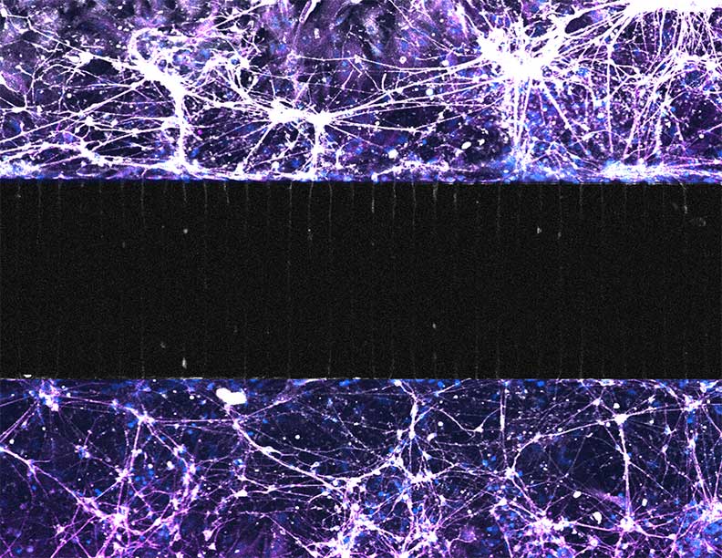
Fluorescence microscopy image of a human iPSC-derived corticospinal tract-on-a-chip with spatially separated cortical (top) and spinal (bottom) compartments that mimic human anatomy. Neurons are shown in white and astrocytes in purple, and the compartments communicate through corticospinal axons projecting across the intervening microchannels (black region).
Johns Hopkins neurologist Nicholas Maragakis and a team of neurologists and bioengineers have created a human-derived corticospinal cell system, for use in researching neurodegenerative diseases like ALS and primary progressive multiple sclerosis (PPMS). The findings were published in June in the journal Cell.
“This is a first step in making a corticospinal network that is versatile, that is relatively easy to set up in a laboratory, and that can be broadly applied to neurodegenerative diseases from a variety of different angles,” Maragakis says.
Neurodegenerative disorders are characterized by decay of the neurons connecting the brain and spine along the corticospinal tract. Variations in where the involvement begins and how it spreads are still poorly understood, making treatments less effective than they could be.
“A drug that might be good for one form of ALS may not be good for another,” says Maragakis. “Yet right now we're lumping them all together for ALS as one disease.”
Until recently, researchers have had difficulty representing these differences in laboratory settings because cells derived from flies and other animal models lacked the network activity of human cells, Maragakis says. “We were very interested in making a humanized model that we could manipulate.”
The solution is a cell system derived from the pluripotent cells of healthy humans, with four different types of cells: cortical neurons and astrocytes, and spinal neurons and astrocytes. Unlike previous models, these cells have synaptic connectivity, allowing real-time analysis of corticospinal tract activity as researchers study various interventions.
“It's taken a lot of heavy lifting from people in my laboratory over the last several years to get to this point,” says Maragakis. “There were a lot of moving parts to the study, including getting those nerve cells to grow, and getting them to differentiate into their appropriate cell types.”
“It was really important to get the foundation right, to show that we could establish connectivity between the brain neurons and the spinal neurons, not only anatomically, but electrophysiologically as well.”
The effort took between three and five years, he says, with “a lot of trial and error.” The team includes neurologists Andriana Charalampopoulou, Arens Taga, Khalil Rust, Evelyn Luciani, Katherine Marshall, Elliot Montgomery, Anuradha Mansinghka, Richa Singh, Arun Venkatesan and Christa Whelan Habela; and bioengineers Yang Zhao, Christine O’Keefe and Tza-Huei Wang.
The breakthrough allows researchers to manipulate the cells for research, eventually using cells from ALS patients with specific mutations or clinical manifestations.
“That’s really where the field is going, towards this precision medicine approach, both as we target genetic forms of the disease, but also as we try to understand why specific cells respond differently in these patients,” says Maragakis.
His lab is already using the model to study how ALS passes from the brain to the spinal cord. “Even more exciting is that we’ve had a lot of interest from other ALS investigators, who want to use our system to ask specific questions,” he says. “That kind of collaboration is really the intent of this model system, not keeping it for ourselves.”
For Clinicians Clinical Connection
Clinicians, discover the latest in research and clinical innovation from Johns Hopkins experts. Access educational videos, articles, CME courses and other resources from our world-renowned institution.