With New Technology, Endovascular Surgery Leaps Forward at Johns Hopkins
Key Points
- The Johns Hopkins Hospital is the first in the mid-Atlantic region to adopt fiber optical vessel navigation technology, which uses radio frequency for visualization of blood vessels during procedures.
- Images are rendered in 3D, improving accuracy and reducing procedure time.
- Radiation exposure to the patient and treatment team is dramatically reduced.
The Johns Hopkins Hospital is the first in the mid-Atlantic region and one of fewer than 10 medical centers nationwide to use an imaging device that augments the guidance of catheters during endovascular procedures and dramatically reduces radiation exposure.
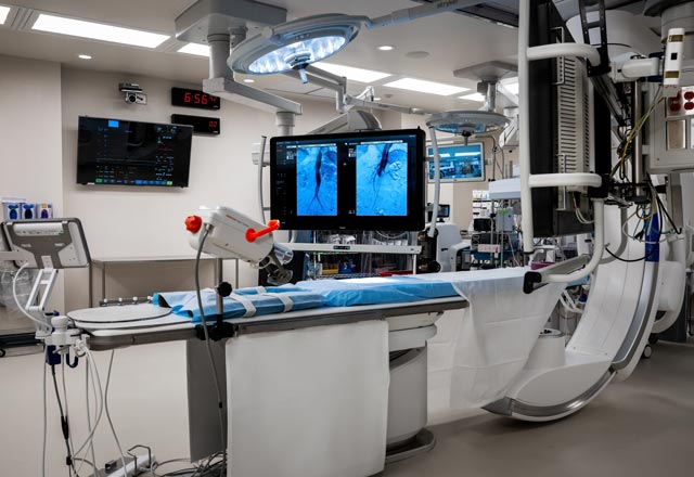
The hospital acquired the fiber-optic vessel navigation technology in January as part of a comprehensive upgrade to its hybrid endovascular operating room, which hosts both open and endovascular surgeries for patients with emergent and long-term aortic issues.
"It’s being used for endovascular procedures including aneurysm repairs, stents and ballooning of arteries," says James Black, chief of the Division of Vascular Surgery and Endovascular Therapy. “Essentially every vascular patient can benefit,” he says.
The new equipment, now in a larger room, uses radio frequencies to provide live, in-body imaging in three dimensions, which is then projected on 110-inch monitors that surgeons view to guide catheters for diagnosis and treatment. (The previous monitors, pre-upgrade, were just 17 to 20 inches, says Black.) It also allows fusion imaging with prior CT scans to greatly increase accuracy in treatment of aneurysms and aortic dissections.
Previously, these procedures relied on X-ray machines for visual guidance, which exposed patients — and to a lesser extent, others in the operating room — to radiation.
"The visualization is much, much better. We have better detail, better understanding of our patient's anatomy."
James Black, chief of the Division of Vascular Surgery and Endovascular Therapy
Black continues, "We get a 3D model, using a catheter and wire combination, that senses its position in three-dimensional space inside the patient and generates a cartoon-like image that allows us to catheterize vessels and complete procedures with absolutely zero radiation exposure to anyone."
The improved imaging means the procedures typically take less time — a particular boon to patients who don’t tolerate anesthesia well, he says. There’s also less risk of error. “The overlying image that has the aorta and the aneurysm and the branches allows us to align the stent that we’re placing in the patient better, because it takes away a lot of the estimations and assumptions,” he says. “You know exactly where the vessel is running, and where your target areas are.”
He also notes that the technology marrying CAT scans to live images requires smaller doses of contrast dye, reducing risk of future kidney dysfunction. “Those are big wins for everyone,” he says. “It’s of course only a tool, but having the best tool and the best technology available makes the procedures go better for the patient and better for us.”
Medically reviewed by James Black, M.D.
For Clinicians Clinical Connection
Clinicians, discover the latest in research and clinical innovation from Johns Hopkins experts. Access educational videos, articles, CME courses and other resources from our world-renowned institution.