
A Transformative Transplant
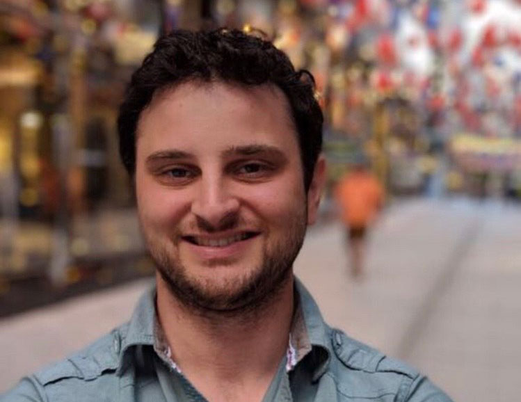
In August 2023, Jonathan Knapp was on an Alaskan cruise with his family when his eyes suddenly became severely painful and sensitive to light, to the point where he couldn’t even tolerate the hallway light peeking in from under the door to his cabin.
Earlier that year, Knapp had developed a fungal infection in both of his eyes. As someone previously diagnosed with keratoconus — a condition that thins the cornea, the clear outer covering of the eye — he was more susceptible to such infections. He was prescribed intensive antifungal eyedrops that he needed to take hourly. Just prior to the cruise, he was told that the fungus had cleared, and was switched to immunosuppressive eyedrops to treat scarring that resulted from the infection. But that information turned out to be wrong; the fungus was still there. When it reemerged in his already compromised eyes, the pain was worse than before.
Upon returning home from his trip, Knapp saw cornea specialist Samuel Yiu, M.D., Ph.D., of the Wilmer Eye Institute, Johns Hopkins Medicine. Yiu referred Knapp to fellow cornea specialist Nakul Shekhawat, M.D., M.P.H., Sc.M., whose expertise is in complex and severe corneal cases. Shekhawat, the Stephen F Raab and Mariellen Brickley-Raab Rising Professor of Ophthalmology, saw that Knapp’s corneas were opaque with dense pus.
“Jonathan’s situation was about as severe as it gets,” Shekhawat says. “Fungal keratitis is a potentially devastating, blinding disease that usually only occurs in one eye. With both eyes involved, he had a complete loss of ability to see.”
When evaluating Knapp, the central question, Shekhawat says, was whether to continue aggressive medication alone, such as taking hourly antifungal eye drops, or to move quickly to surgery. Delayed or inadequate treatment could lead to the infection spreading deeper, and even loss of the eye.
Shekhawat used a state-of-the-art anterior segment optical coherence tomography scanner to examine Knapp’s corneas and the extent of the fungal infection in each eye. This high-resolution imaging quantifies the depth of fungal infiltration, an objective measurement that can predict whether an infection will respond to medications or require surgical intervention. The scans revealed that the left eye’s infection would likely respond to medication, while the right eye showed fungal invasion into deeper layers beyond the reach of medications. Shekhawat concluded that medication could treat the left eye, but emergency surgery was needed for the right eye to save it from permanent damage.
That same night, Knapp underwent a therapeutic penetrating keratoplasty — also known as a full thickness corneal transplant — for his right eye, performed under the direction of Allen Eghrari, M.D., M.P.H.
The infected cornea was removed, and a clear donor cornea was secured in its place with 16 stitches. Shekhawat oversaw perioperative management, tailoring antifungal and immunosuppressive therapy to maximize the likelihood of infection eradication and corneal transplant survival, based on protocols developed through years of experience and his team's ongoing outcomes research.
For the left eye, Shekhawat relied on gold standard randomized clinical trial data to determine the optimal antifungal medication Knapp needed to eliminate the fungal infection for good.
Knapp, now 35, says his vision quickly improved and pain subsided within two weeks. He can now perform daily tasks without assistance and uses contact lenses when detailed focus is needed. He says he is grateful for the Wilmer staff and for his mother, who accompanied him to appointments and assisted with his care at home.
“Everyone was extraordinary,” he says. “They always kept me informed of what was going on. They didn’t sugarcoat anything, but at the same time they provided feedback on all the options that were available. I always felt that I was in great hands.”
Knapp’s two eyes, each treated differently but successfully, offer a rare side-by-side comparison of surgical versus medical management of corneal infections in the same patient. For Shekhawat, Knapp’s case crystallized questions he had been grappling with across hundreds of similar patients: When does medication become insufficient, and when is surgery necessary to successfully treat the infection and save the eye?
To address this challenge, Shekhawat launched a multicenter study spanning five eye hospitals around the world. The global consortium has collected data and images from more than 6,000 patients with corneal infections and more than 200 patients who underwent therapeutic keratoplasty.
The goal, Shekhawat says, is to identify which patients with severe corneal infections benefit most from transplantation, which patients may do well with medication alone, and which preoperative factors predict good or poor outcomes — ultimately improving decision‑making and outcomes for patients worldwide.
Insights from this effort are already informing care at Wilmer, including five recent fungal infection cases requiring therapeutic transplants over the past few months. “We gain intuitions from years of clinical experience, but that kind of pattern recognition isn’t always easy to teach or transfer,” Shekhawat says. “Our goal is to translate that expertise into objective evidence that doctors everywhere can use to help their own patients.”