Visionary in Medical Illustration
Annette Smith Burgess was recruited by Dr. William Holland Wilmer in 1926 to become the institute’s first ophthalmic illustrator, a role she held for more than three decades.
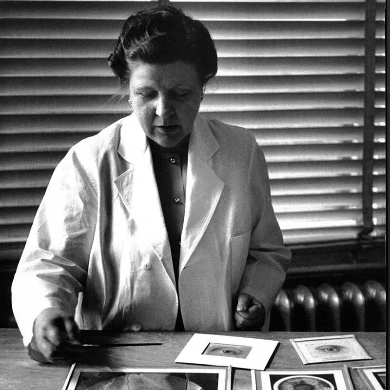
Annette Smith Burgess, from an article in the 1953 edition of The Johns Hopkins Magazine titled “Art in Medicine.” Photo credit: Werner Wolff
Before cameras could capture the finest details of the inside of the eye, the responsibility of recording the intricacies of ocular anatomy and pathology fell to artists whose tools were not lenses and sensors, but pencils, brushes and extraordinary powers of observation. This rare combination was embodied by Annette Smith Burgess, who could document the minutiae of the eye to such levels that her sketches and paintings have remained educational tools nearly a century after they were created.
Burgess was recruited by Dr. William Holland Wilmer in 1926 to become the institute’s first ophthalmic illustrator, a role she held for more than three decades. She had previously studied under Max Brödel, the father of medical illustration and founder of the Department of Art as Applied to Medicine at Johns Hopkins Medicine.
“Mrs. Burgess was unique in the world,” says Morton F. Goldberg, M.D., the Joseph E. Green Professor of Ophthalmology and Director Emeritus of the Wilmer Eye Institute. “The tiny things she painted were absolutely accurate and precise. Nobody else could do that then; nobody else could do that now.”
Without digital photography or advanced scanning technologies, capturing the internal structures of the eye was a formidable challenge. Observers were often allowed only brief glimpses through the ophthalmoscope. Burgess would spend hours carefully observing the eyes of Wilmer’s patients — both normal and abnormal features — for just a few seconds at a time. She memorized the details, and then took what she saw to paper.
“She had the most amazing visual memory,” Goldberg says. “That’s why I admire her so much. She was the world’s expert at observing, understanding, remembering, painting and drawing over and over until she finished the job.”
These illustrations served multiple purposes: They helped explain conditions to patients, educated medical students and doctors, and provided a clinical record to track patient progress over time. Many of her paintings were featured in Atlas Fundus Oculi, William Holland Wilmer’s 1934 color atlas of the retina and choroid, which remains one of the most admired and authoritative texts on the subject today.
At Wilmer, Burgess mastered the use of the direct ophthalmoscope to visualize the retina and choroid — structures that make up the fundus of the eye — as well as slit-lamp microscopy to examine the anterior chamber. This diagnostic tool uses a specialized microscope (the slit lamp) to examine the detailed structures of the eye — particularly the front part, including the cornea, iris and lens — by directing a narrow beam of light into the eye, allowing for close inspection and diagnosis of various eye conditions.
To draw the external eye, Burgess used nothing more than a bright flashlight to illuminate the eyelids, eyebrows, eyelashes and the colored and white portions of the eye. To capture the retina, she guided the patient's gaze through 12 meridians to carefully observe and capture every nuance of the retina's structure, ensuring accuracy in color, shape and location. Her work showcases not only the complexity of the eye but also the exceptional skill and dedication required to illustrate its hidden intricacies.
Burgess’s images have withstood the test of time and remain vivid illustrations of ocular disease, says James Handa, M.D., chief of Wilmer’s Retina Division and the Robert Bond Welch, M.D. Professor of Ophthalmology. He says her work is remarkable both in its accuracy for capturing ocular anatomy and disease, and for how challenging this endeavor was.
“I think any ophthalmologist who sees her work will appreciate how amazing the images are, how they accurately depict disease and eye anatomy, and how much she contributed to ophthalmology,” he says. “For me, seeing her work makes me really proud to be a part, albeit minuscule compared to her, of Wilmer.”
The Legacy of Annette Smith Burgess
Annette Smith Burgess left Wilmer more than 800 original paintings — many unpublished — which the institute has preserved since the 1920s. A new exhibit, created with the Department of Art as Applied to Medicine at Johns Hopkins, showcases select works and honors Burgess’s legacy.
Located on the fourth floor of Wilmer’s Maumenee Building, “A Century of Wilmer Illustrations: The Legacy of Annette Burgess,” features two original watercolor paintings and 14 color reproductions from Atlas Fundus Oculi, William Holland Wilmer’s 1934 color atlas of the retina and choroid.
The exhibit also has about 70 previously unpublished Burgess paintings on display, as well as 16 works by other Wilmer illustrators. Artifacts related to the creation and publication of the atlas at the nearby Hoen lithography company are also featured.
See below for a sampling of images by Annette Smith Burgess.


For Clinicians Clinical Connection
Clinicians, discover the latest in research and clinical innovation from Johns Hopkins experts. Access educational videos, articles, CME courses and other resources from our world-renowned institution.