Evolution of Excellence
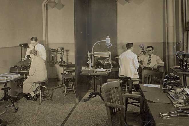
Clinic exam room, 1929
For the past century, researchers at the Wilmer Eye Institute, Johns Hopkins Medicine, have driven groundbreaking advances in ophthalmology, including pioneering earlier diagnoses, revolutionizing treatments and transforming patient outcomes.
Some of the most pivotal breakthroughs include: In 1979, Harry Quigley, M.D., made a sight-saving discovery when he learned that nerve cells are the key factors in earlier diagnoses of glaucoma. More recently, Peter Campochiaro, M.D., led a study that determined the efficacy of an implant that provides continuous treatment for wet age-related macular degeneration. And Divya Srikumaran, M.D., has led studies using big data to help diagnose corneal diseases and assess complication risk after corneal transplants. That work is still evolving — and advancement in artificial intelligence (AI) offers exciting possibilities for the future.
In the stories that follow, we shed new light on each of these game-changing contributions.
EARLIER DIAGNOSES FOR PATIENTS WITH GLAUCOMA
 Harry Quigley
By the mid-1970s, experts knew that glaucoma is a chronic disease that causes increased eye pressure, which ultimately leads to vision loss and blindness. However, diagnosing the disease was a challenge, because early-stage glaucoma does not cause symptoms and vision loss is gradual. Ophthalmologists used an ophthalmoscope to look for a cupped appearance in the center of the optic nerve disc, but this indicated an advanced stage of glaucoma. As a result, patients often were not diagnosed until their vision was irreversibly affected.
That changed in 1979, with publication of a landmark study led by Harry Quigley, M.D., now the A. Edward Maumenee Professor of Ophthalmology, which provided new insight into how early glaucoma was evading diagnostic methods. In collaboration with Wilmer pathologist W. Richard Green, Quigley authored “The Histology of Human Glaucoma Cupping and Optic Nerve Damage: Clinicopathologic Correlation in 21 Eyes,” which the journal Ophthalmology honored as one of the seven most influential papers in its history. For Quigley, this was the beginning of decades of glaucoma research — research that has contributed to earlier diagnosis and timelier, sight-saving treatment.
Quigley notes that when he began his investigations in the mid-1970s, research on the mechanisms of glaucoma centered on studying eyes from pathology labs that had been removed after becoming painful and the patient was blind in that eye, but not from people with typical primary glaucoma.
“None of the research was meaningful in terms of actually understanding glaucoma, because we weren’t collecting eyes from people who had the typical disease at its various stages,” Quigley says. Few researchers were focused on understanding the time course of glaucoma blindness.
Quigley became one of them.
In 1975, Quigley, who had recently completed his residency at Wilmer, started a two-year glaucoma research fellowship at the Bascom Palmer Eye Institute in Miami, at the encouragement of then Wilmer director Edward Maumenee. At Bascom Palmer, while researching glaucoma on animal models, Quigley gained access to a valuable resource: an eye from a patient who had glaucoma for many years, donated by his family. This was the first time Quigley was able to investigate the effects of primary glaucoma on a human eye for which clinical findings during the donor’s life were available. This eye became part of his landmark study.
After his fellowship, Quigley returned to Wilmer, where he collaborated with Green by gathering eyes to study after autopsies and from eye banks and surgical departments. When Quigley had 21 eyes from patients who had chronic glaucoma, he looked at the structure of the eyes’ cells and tissues using standard light microscopy and electron microscopy along with the donors’ clinical records — a method known as clinical-pathological correlation.
From this, Quigley and Green learned that the loss of retinal ganglion cells — nerve cells that transfer data from the retina to the brain through the optic nerve — was occurring much earlier than ophthalmologists believed. Further, the reason for the cupped appearance in the center of the optic nerve disc was that ganglion cells were dying. “We essentially redefined the initiation of the disease to an earlier phase,” Quigley says.
During the next decade, he and Wilmer colleagues Neil Miller, M.D., who would lead Wilmer’s neuro-ophthalmology division, and Alfred Sommer, M.D., M.H.S., founding director of the Dana Center for Preventive Ophthalmology at Wilmer, pursued the theory that the number of dying cells could be measured by looking at the nerve fiber layer — an area in the retina where nerve cell fibers pass down toward the optic nerve. That finding ultimately led to daily use in every ophthalmology office of instruments and techniques, including optical coherence tomography (OCT), to quantitatively measure the number of nerve fibers remaining in the retina to stage glaucoma and follow its course.
Timely detection of the disease is key. A preliminary nerve cell loss of 30%–40% indicates that glaucoma has started. Finding this loss leads to earlier treatments than had previously been possible. Vision can be preserved if a patient is treated with eye drops, lasers or surgery during the early stages of glaucoma. “That’s where this paper went,” Quigley says.
WET AMD: BRINGING A NEW TREATMENT TO MARKET
 Peter Campochiaro
Age-related macular degeneration (AMD) damages the macula in the center of the retina, leading to vision loss that affects the ability to recognize faces and perform everyday activities such as driving, reading and cooking. In the United States, 11 million people primarily over the age of 60 have either wet or dry AMD.
Wet AMD — which accounts for 90% of AMD cases that lead to legal blindness — occurs when changes in dry AMD, such as deposits under the retina, are accompanied by increased production of vascular endothelial growth factor (VEGF) by retinal cells. VEGF is a protein that causes abnormal blood vessels to grow under the retina and leak fluid into the macula, which reduces vision.
Since the early 2000s, treatment for wet AMD has included frequent injections of antibodies or other proteins that block VEGF. The injections are effective at preserving vision, but the proteins exit the eye within a short amount of time. Patients must receive repeated injections, often every four to six weeks, to keep fluid from re-accumulating and maintain their vision — a challenging schedule to sustain, notes Peter Campochiaro, M.D., the George S. and Dolores D. Eccles Professor of Ophthalmology.
“It can be difficult for patients to get back to the doctor’s office that frequently,” Campochiaro says. “But if treatments aren’t given in a timely manner, the leakage comes back and can produce scarring, and vision can gradually decrease.”
An implant developed by Forsight Vision Care and Genentech was designed to address this challenge, explains Campochiaro, an adviser to Genentech. The implant contains ranibizumab, an antibody fragment that is slowly released into the vitreous cavity in front of the retina and that blocks surplus VEGF. The implant, which is inserted during a 15-minute surgical procedure, spans from under the conjunctiva to the inside of the eye. Patients only need to have the implant refilled every six months — a much more manageable schedule.
Campochiaro helped bring the implant to market by leading a phase II clinical trial that determined its efficacy and safety. The Food and Drug Administration approved the implant in 2021.
For the phase II study, 220 people received the implant with one of three doses of the antibody fragment. The study looked at the length of time it took for fluid to build up in the macula again with each dose. In more than 95% of the group that had the highest dose, the fluid started to come back in six months. This dose of antibody fragment is used now in the implant.
Today, the implant is widely available. It offers the first successful approach to sustained, long-term suppression of VEGF for patients with wet AMD, one that has stimulated the development of other long-term treatments, Campochiaro notes. For example, gene therapy, which is currently in clinical trials, involves the one-time injection of a gene under the retina, which then causes the retinal cells to continuously make a protein that blocks VEGF.
“The implant was the beginning of this new wave of long-term treatments for wet age-related macular degeneration that is improving outcomes and reducing burden for this disease,” says Campochiaro.
HARNESSING BIG DATA AND AI TO IMPROVE OUTCOMES FOR CORNEAL TRANSPLANTS
 Divya Srikumaran
During the first half of the 1900s, patients who needed surgery due to a damaged cornea had a full-thickness corneal transplant, during which the entire cornea was replaced with healthy donor tissue. But this procedure requires long recovery periods, and graft rejection occurs in up to 30% of cases. About 70 years ago, ophthalmologists started to consider replacing only the diseased portion of the cornea. Charles Tillet, then a Wilmer resident, performed the first partial corneal transplant in 1956. This was groundbreaking, and it took another 40 years for use of partial corneal transplants such as DSEK (Descemet stripping endothelial keratoplasty) and DMEK (Descemet membrane endothelial keratoplasty) to gain traction.
These procedures are now commonly performed, and researchers at Wilmer are continuing to advance care for patients who need corneal transplants by leveraging big data and AI to diagnose corneal diseases and assess patients’ risk of post-transplant complications in order to avoid corneal rejection and other complications. The risk of graft rejection at any stage after a corneal transplant is about 18%–21%.
“In a randomized controlled trial, it takes a while to get the data and then you’re limited as to how many variables you’re able to collect,” says Divya Srikumaran, M.D., the Walter J. Stark, M.D., Professor of Ophthalmology and chief of the Division of Cornea, Cataract and External Disease. “Big data and AI can help supplement the analyses because you can use information on patients from a larger range of practice settings and more diverse patient populations.”
For her research on corneal transplants, Srikumaran uses big data — complex data sets that can include text, images and graphs — from sources such as the American Academy of Ophthalmology’s Intelligent Research in Sight (IRIS) registry to look for insights into the factors involved in successful corneal transplants. These factors could include the types of donor tissue used and the surgeons’ experience level. The IRIS registry contains data on more than 70 million patients — representing about 21% of the U.S. population — from more than 15,000 ophthalmologists and 2,900 optometrists.
Using this data, AI algorithms can then be created to help with diagnosis of corneal diseases. Keratoconus, which is characterized by a cone shape on the cornea that leads to blurred vision and requires a corneal transplant in up to 20% of cases, has traditionally been diagnosed by ophthalmologists who look for the cone shape on images of the cornea. But the images can be hard to interpret and there is no consensus on the criteria for diagnosis.
“There’s a lot of ‘noise,’ and sometimes we can’t ascertain the diagnosis or confirm if there is progression,” Srikumaran says. “But with AI, we would be able to have a computer help us more accurately determine which patients have keratoconus and are progressing so we can tailor our therapy and follow up accordingly.”
AI can also be helpful in assessing the risk of graft rejection (when the body recognizes the graft as a foreign entity and attacks it). Physical symptoms usually indicate that a donor cornea is close to rejection, including swelling of the cornea, redness in the eye and lung sensitivity. But AI could make this determination before the physical symptoms appear by sifting through detailed images of the cornea that display the endothelial cells. A reduction in these cells indicates a graft might be about to fail.
“Perhaps then we could up their steroids or treat them before the acute rejection happens and the patient becomes symptomatic,” Srikumaran says, adding that rejection could be reversed.
Srikumaran sees AI becoming an assistive tool for ophthalmologists that supports their diagnostic and follow-up approaches and allows them to provide better patient care. AI has already been used to optimize intraocular lens selection for cataract surgery, which is the most common surgery performed in the U.S. It will also help expand access to care in rural areas of the U.S. and globally, she says.
“AI may be used in low-resource settings to screen populations of patients and target who needs to be referred to an ophthalmologist for a higher level of care,” Srikumaran says. “There are so many ways that AI can help us.”