A Pathology Revolution, One Slide at a Time
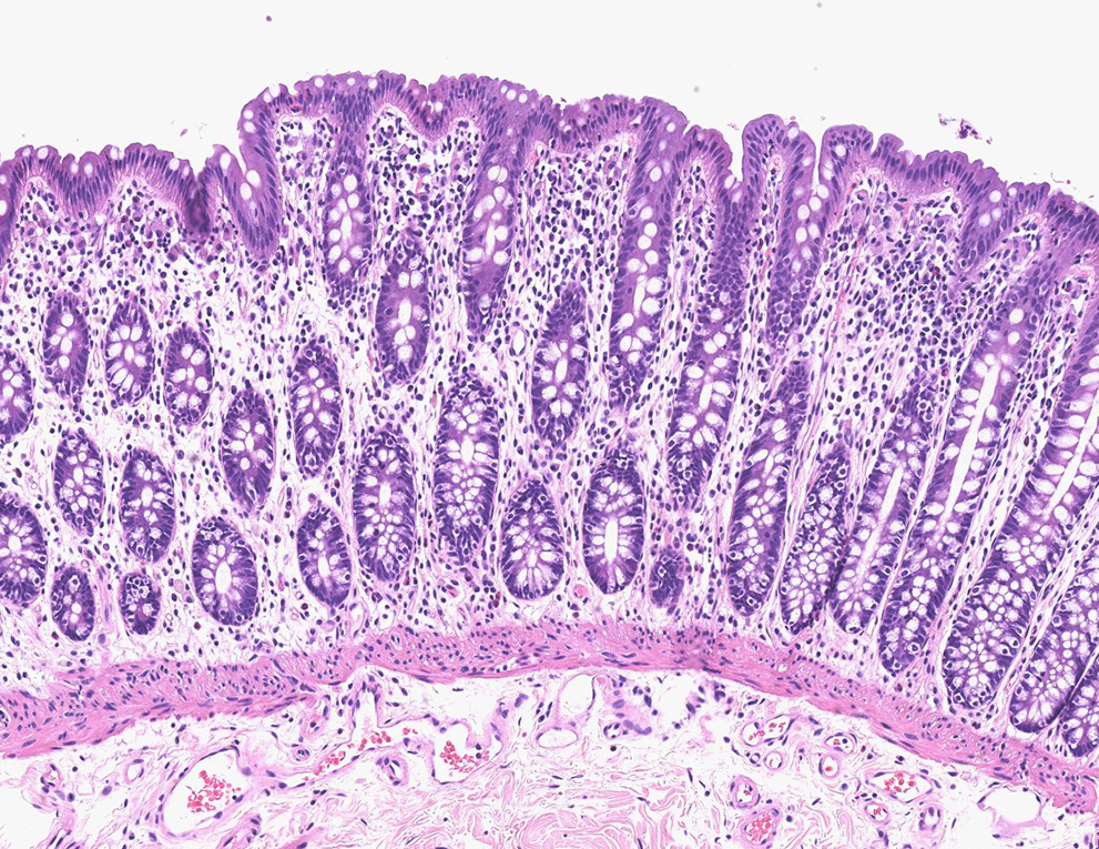
An up-close look at gastrointestinal mucosa
Every slide tells a story.
Assistant pathology professor Ashley Kiemen holds one up, peering at a sliver of tissue, preserved and positioned decades ago between two slim panes of glass. In the slide, she sees a snapshot of a Johns Hopkins patient with a certain type of pancreatic cancer.
The slide captures a single moment in time, perhaps when the cancer was diagnosed, or possibly after a round or two of chemotherapy. It also captures a single place, a location in a person’s pancreas that may or may not be cancerous.
What it doesn’t do, however, is pinpoint the very first signs of disease, analyze how the patient responded to treatment, or add to a dataset that could revolutionize understanding and treatment of pancreatic neuroendocrine tumors.
For all that to happen, Kiemen and her students are digitizing that slide and thousands more, as part of a sweeping movement that is transforming medical research, care and education at Johns Hopkins and elsewhere.
About two years ago, Johns Hopkins Health System clinicians began digitizing patient slides as a matter of routine, and storing the information in patients’ electronic medical records, explains Ralph Hruban, director of the Department of Pathology.
That amounts to about 10,000 slides each month from patients treated at Johns Hopkins, as well as those who bring biopsy materials in for a second opinion, he says.
Yet it’s just a fraction of the millions of Johns Hopkins slides available for digitization, most stored in an off-site facility and representing decades of patient care and second opinions.
“The potential is enormous,” says Hruban.
Researchers such as Kiemen are chipping away at the collection as they digitize specific datasets for their research.
Kiemen, her students, and pathology fellows are scanning about 25,000 slides from Johns Hopkins Hospital and Johns Hopkins Bayview Medical Center patients since the 1990s to create a digital blueprint of pancreatic neuroendocrine tumor cells. There could be five to 25 slides per patient, created over the course of treatment, Kiemen says.

The process, performed in a cluttered, computer-filled room in the pathology building, is time consuming. Each slide has to be examined for damage and linked to patient records, which are sometimes as rudimentary as a photocopy of handwritten clinical notes. If the slides are scanned for research, the files are de-identified.
Kiemen and her team then use CODA, a deep learning tissue mapping platform they developed, to create three-dimensional models of healthy and cancer-containing pancreas specimens, showing the tissues at subcellular resolution.
“This will give us a sense of what the tissue looked like in thousands of different patients with the same disease,” says Kiemen. With that information, clinicians can better diagnose cancers in their earliest stages, and researchers will gain understanding of how these cancerous cells behave. Also, educators will have new tools to aid student learning.
“There is so much information in the slides,” says Alexander Baras, director of pathology informatics. He is leading the digitization effort.
Baras says digital pathology has gained traction in recent years, with scanners that can digitize many slides at a time and cloud storage that has become inexpensive and widely available. (The Johns Hopkins digital pathology public repository is one of several that allows researchers to share their images.)
“Pathology has a long history of embracing automation and technology,” he says. “It’s going to make us more efficient, it’s going to make us more accurate and it will eventually uncover new features of a disease.”
Going forward, it’s clear that artificial intelligence will play a growing role in analyzing digitized tissue information, providing better diagnoses and turbocharging research aimed at understanding diseases.
“It’s this inflection point that is exciting, this revolution that’s occurring,” says Hruban.
With additional reporting by Katie Pearce.