Craniosynostosis
When an infant's skull bones fuse together too early, it can create an irregular head shape — a condition called craniosynostosis.
What You Need to Know
- Craniosynostosis is common; it occurs in 1 out of 2,200 live births.
- The condition affects males slightly more often than females.
- A noticeable change to a baby’s head shape soon after birth is a sign of this condition.
- Craniosynostosis most often occurs by chance, but it can be inherited.
What is craniosynostosis?
Craniosynostosis is a congenital (present at birth) condition in which the flexible joints between the bones of the skull close too early, causing problems with brain and skull growth. These joints, called sutures, are present in fetuses to allow for the skull to fit through the birth canal. As newborns grow and develop, the sutures gradually close, forming a solid piece of bone — a process called synostosis. Early closure of the sutures may cause pressure inside the head to increase and the skull or facial bones to become asymmetrical or misshapen.
Craniosynostosis occurs in 1 out of 2,200 live births, and affects males slightly more often than females. Several treatments exist today to safely and effectively treat craniosynostosis.

Craniosynostosis Overview from Johns Hopkins All Children’s Hospital
Webinar: Understanding and Treating Craniosynostosis

Craniosynostosis Causes
Most cases of craniosynostosis affect only one of the sutures. In these cases, the cause is usually unknown and the chance of it happening again in another child born to the same parents is low.
In more complex cases, craniosynostosis may affect multiple sutures of the skull, other parts of the face, and parts of the body outside of the head and neck, such as hands, feet and body organs. Complex cases like this are called syndromic craniosynostosis because the condition is likely caused by a genetic syndrome.
Craniosynostosis Symptoms
Difference in Head Shape
In infants with craniosynostosis affecting a single suture, the most common symptom is an atypical shape of the head. The closure of different sutures results in specific changes in the head shape, which include:
- Bulging of the forehead on one side
- Bulging of the forehead and back of the head
- An unusually long head
- An irregularly shaped face and skull (an even rarer condition that occurs when two sutures fuse)
The head changes shape when a suture closes early because the brain starts growing parallel to the suture. Doctors call this Virchow’s rule. For example, if the suture that divides the skull into left and right halves fuses early, the brain grows lengthwise (parallel to the suture), causing bulging in the forehead and in the back of the head.
Elevated Intracranial Pressure
In 10%–15% of craniosynostosis cases, only one suture fuses, and the child may develop increased brain pressure as they grow. In more complex, syndromic cases of craniosynostosis, the risk of elevated pressure is higher. Surgery can help prevent this.
Signs of elevated intracranial pressure may include:
- A full or bulging fontanelle (soft spot located on the top of the head)
- Sleepiness (or being less alert than usual)
- Very noticeable scalp veins
- Increased irritability
- High-pitched cry
- Poor feeding
- Projectile vomiting
- Increasing head circumference
- Developmental delays
The symptoms of craniosynostosis may resemble other conditions or medical problems, so parents should work with the child’s physician to clarify the diagnosis.
Is craniosynostosis painful?
In many cases, craniosynostosis is not hurting the child and is largely a cosmetic problem. However, it may be severe enough to need corrective surgery.
Craniosynostosis: Adaya's Story

Adaya was 3 weeks old when she was diagnosed with craniosynostosis. At 9 weeks old, she had a successful minimally invasive surgery at Johns Hopkins All Children's Hospital.
Types of Craniosynostosis
Sagittal Craniosynostosis (Scaphocephaly)
The sagittal suture runs across the middle of the skull, from the front to the back. Early fusion of the sagittal suture causes the skull to become long from front to back and narrow from ear to ear. This head shape is called scaphocephaly. Sagittal craniosynostosis is the most common type of craniosynostosis, accounting for about half of all cases.

Coronal Craniosynostosis
The right and left coronal sutures run across the top of the baby’s head from ear to ear. If the sutures fuse early, this is called coronal synostosis, and it causes the bones of the forehead and brow to stop growing.
- Bicoronal or bilateral craniosynostosis: An early closure of the coronal suture on both sides that prevents the front of the skull from growing. This results in a skull that is short (from front to back), wide and tall, with a flat forehead and flat back of the head, called brachycephaly.
- Unicoronal or unilateral craniosynostosis: An early closure of the coronal suture on one side. This results in flattening of the forehead and the brow on the affected side, while on the opposite side the forehead tends to bulge. The eye on the affected side may also have a different shape, and the back of the head may become flat. The resulting head shape is called anterior plagiocephaly.

Metopic Craniosynostosis (Trigonocephaly)
The metopic suture runs from the top of the head down the middle of the forehead, toward the nose. Early closure of this suture may cause a prominent ridge running down the forehead. The resulting head shape is called trigonocephaly (triangular shape). In rare cases of metopic craniosynostosis, the forehead looks pointed like a triangle, with closely placed eyes.
A ridge in the forehead is not always suspicious. Also, the metopic suture is one of the earliest to close in healthy babies, so determining if it closed due to craniosynostosis can be challenging. A craniofacial surgeon or neurosurgeon can tell the difference between a normal ridge and craniosynostosis, and can recommend appropriate treatment.

Lambdoid Craniosynostosis
The lambdoid suture runs across the very back part of the skull, defining the base of the skull. The lambdoid suture may close early on one or both sides:
- Unilambdoid craniosynostosis: An early closure of the lambdoid suture on one side. This causes flatness of the back of the head, tilting of the head and shifting of the ears. The resulting head shape is called synostotic posterior plagiocephaly.
- Bilambdoid craniosynostosis: An early closure of the lambdoid suture on both sides. This may lead to a skull that is unusually wide — a shape called posterior brachycephaly.
Lambdoid craniosynostosis is the least common form of single-suture craniosynostosis. It occurs in only 1% of craniosynostosis cases. A similar flattening of the back of the head can happen when babies lie on the back for a long period of time. This is called positional or deformational plagiocephaly, and can occur in 1 in every 3 infants. A craniofacial expert will be able to tell the difference between this condition and craniosynostosis.
Multi-Suture Craniosynostosis
In rare cases, multiple sutures may close early at the same time. The affected sutures may include:
- Sagittal and metopic sutures: The resulting head shape is very narrow and long, called scaphocephaly.
- Coronal, lambdoid and metopic sutures: The resulting head shape is called Kleeblattschädel, or cloverleaf. This is the most severe form of craniosynostosis.
- Sagittal, coronal and lambdoid sutures: The resulting head shape is called acrocephaly, or tower skull, referring to an unusually tall head.
- Sagittal, bicoronal and metopic sutures: The resulting head shape is short, wide and may be pointy at the top.
When multiple sutures fuse early, the cause could be related to an underlying genetic syndrome. Read more about syndromic craniosynostosis.
Craniosynostosis Diagnosis
Trained craniofacial surgeons and neurosurgeons can tell the difference between craniosynostosis and other causes of head differences.
Although craniosynostosis is a congenital condition, it is not always diagnosed at birth. Sometimes, it is discovered during a routine physical exam in the first year of the baby’s life.
Diagnosis efforts may include:
- A complete prenatal and birth history, including family history of craniosynostosis or other head or face anomalies. The doctor may also ask about developmental milestones, because craniosynostosis can be associated with other neuromuscular disorders. If developmental delays are present, they may prompt follow-up to discover underlying problems.
- Physical exam. During the exam, the doctor may check for:
- The shape of the fontanelle (the spot at the top of the skull where several bones meet)
- Ridges between the bones of the skull
- Position of the ears
- Overall shape of the head and facial features
- Measurements of the head circumference
- Craniosynostosis can often be diagnosed by medical history and physical exam alone. If needed, the doctor may recommend imaging tests such as low-dose head CT scan, MRI or ultrasound.
- If the doctor suspects syndromic craniosynostosis, they may also recommend genetic testing to help identify specific genetic syndromes.
Can craniosynostosis be diagnosed with an ultrasound during pregnancy?
Rarely. There are times when a sonographer might notice something about the baby’s head shape while the baby is still in the womb. However, ultrasound technology is not refined enough to allow for a diagnosis before a baby is born.
Craniosynostosis Treatment
Early diagnosis and consultation with a specialist are important for successfully treating craniosynostosis. The child’s doctor will work with the child’s parents or guardian to recommend treatment options based on:
- The child’s age, overall health and medical history
- Severity of the craniosynostosis
- Type of craniosynostosis (which sutures are involved)
- The child’s tolerance for specific medications, procedures or therapies
- Expected progression of the condition
Treatment options may include:
- Surgery: Most children need surgery (sometimes multiple surgeries) to correct the head shape, relieve the pressure inside the head and address any other issues, such as asymmetrical face features. Surgery is often recommended before the child is 1 year old, because the bones are still soft and easy to work with. In cases of severe craniosynostosis, the doctor may recommend surgery as early as 1 month of age.
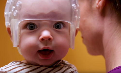
- Helmet therapy: The child will wear a special helmet to help reshape the head. Helmets may be used as part of a treatment after some types of craniosynostosis surgery.
After getting treatment for craniosynostosis, children should have regular checkups with their doctor. This helps monitor the healing process and check for developmental milestones.
Craniosynostosis: Fitz’s Story
When Fitz was born, it was obvious that his skull was misshapen. By 5 weeks old, Fitz had been diagnosed with craniosynostosis. His skull had fused early and was constricting his brain growth.
What happens if craniosynostosis is left untreated?
If left untreated, craniosynostosis and the resulting increased intracranial pressure can lead to:
- Headaches
- Seizures
- Issues with vision (including blindness), hearing and speech
- Delays in development
- Growth restriction and damage to the brain
The most severe untreated cases with intracranial pressure can result in death.
Prognosis and Life Expectancy for Children and Adults with Craniosynostosis
Craniosynostosis can affect a child’s brain and its development, as well as cause cosmetic issues. However, with timely diagnosis and early treatment, the shape of the skull can be improved, reducing pressure on the brain and restoring facial symmetry. This means the child can have a normal life expectancy and good quality of life.
Some children with craniosynostosis may experience developmental delays and intellectual or behavior problems, including issues with learning, speech, language, communication and memory. Researchers are studying whether developmental delays are related to the restricted brain growth, high intercranial pressure, or whether both craniosynostosis and developmental delays are caused by the same genetic anomaly.
Children with some forms of syndromic craniosynostosis may have a higher likelihood of developmental delays and intellectual disability, as well as a shorter lifespan. This is due to additional anomalies caused by the genetic syndrome, which may include neurological issues, breathing problems and heart problems.