Suites and Equipment at the Center for Image-Guided Animal Therapy
The Center for Image-Guided Animal Therapy (CIGAT) is one of the first centers in the world to take equipment, originally designed for humans, that has undergone further modification and organization in order to optimize the results to advance veterinary care. In addition, each veterinary patient benefits from protocols and software developed at the Johns Hopkins University School of Medicine.
CIGAT has an array of minimally-invasive suites available to the veterinary patient allowing more effective diagnostics and therapies. All suites can be incorporated with sterile procedures and include rooms with:
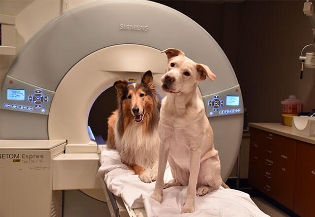
- High-performance, high-field (1.5 Tesla and 3 Tesla) magnetic resonance imaging (MRI) scanners
- Computed tomography (CT) scanners
- Combined Positron Emission Tomography (PET)-CT scanners
- Combined Single Photon Emission Computed Tomography (SPECT)-CT scanners
- Fluoroscopy
- Angiography
- 3-D ultrasound
- Radionuclide imaging scanners (PET and SPECT/CT)
- Radiation Therapy
Each suite is integrated with OR-grade anesthetic medical gases, compatible anesthesia delivery systems, and compatible anesthesia monitoring systems. Fiberoptic communication systems (OptoAcoustics) enable the veterinarians, veterinary technicians and imaging technologists to work seamlessly during MRI procedures.
CIGAT offers the most advanced, minimally invasive diagnostics for animals with disease. The center is designed to create a unique multidisciplinary setting that draws on innovations and diagnostics in musculoskeletal disease, cancer, diseases of the brain, spinal cord, spine, thyroid, lungs, heart, liver, kidney, spleen, pancreas, bladder, prostate, lymphatic system, vascular system, bone and more.
Contact Us
To learn more about CIGAT, send an email to [email protected] or call 410-502-7325