Radiology Exam: Ultrasound Scan
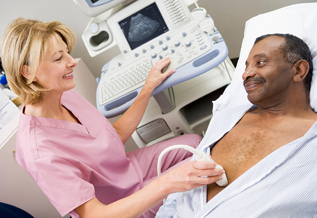
An ultrasound scan is a device that uses high-frequency sound waves to create images of the inside of the body. The scans are used to assess soft tissue structures, including muscles, blood vessels, the heart and various organs.
Technological advancements in the field of ultrasound now include images that can be made in a three-dimensional view (3-D) and/or four-dimensional (4-D) view. A 4-D is a 3-D view that also shows movement.
Ultrasound Scan: What You Need to Know
Common ultrasound procedures are performed to:
- Examine many different parts of the body, including the liver, kidneys, gall bladder, pancreas, breasts, female pelvis, prostate, scrotum, thyroid, heart and the vascular system.
- Evaluate blood flow in a vessel. Doppler ultrasound can determine if there are any blood clots or blockages in the arteries or veins.
- Assess the development and growth of the fetus during pregnancy.
- Guide various types of biopsies and invasive procedures, including thyroid biopsies, liver biopsies, prostate biopsies, kidney transplants and amniocentesis.
Request an Appointment
Schedule by phone
New and existing patients: 443-997-7237

Why Choose Johns Hopkins Radiology for Ultrasound procedures?
Our Physicians
Our diagnostic radiologists have subspecialty training in interpreting ultrasound results to assist in determining the best course of treatment. Our state-of-the-art equipment and technology is combined with providing the highest level of patient care.
Find a Johns Hopkins radiologist who specializes in ultrasound scans.

Our Locations
Ultrasound scans may be done on an outpatient basis or as part of a hospital stay. Ultrasounds are offered at all of our locations. To schedule an exam, call 443-997-7237.
Johns Hopkins Medical Imaging - Bethesda
Johns Hopkins Medical Imaging- Columbia
Johns Hopkins Medical Imaging - Green Spring Station
Johns Hopkins Medical Imaging - White Marsh
The Johns Hopkins Hospital
The Johns Hopkins Outpatient Center
Johns Hopkins Bayview Medical Center