Radiology Exam: Bone Density Scan
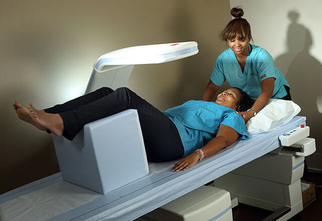
A bone density scan is a diagnostic radiology exam used to measure bone mineral content and mass. This is usually performed with an X-ray, called a DEXA scan, or with a computed tomography (CT) scan of the bones.
Bone density scans can detect the depletion of bone calcium and protein, which increases your bone fracture risk. A bone density scan typically measures the density of the spine, pelvis, lower arm and thigh.
Bone Density Scan: What You Need to Know
- Bone density scans are used primarily to diagnose osteoporosis, monitor the effects of treatment, or determine your rate of bone loss and fracture risk.
- A DEXA scan produces more detailed images of bones than a standard X-ray. This helps to show decreased bone mass and identify fragile bones before they break.
- To prepare for your bone density scan, do not take calcium supplements the day of your exam.
-
There are some portable DEXA or ultrasound units used for general screenings that examine the heel, lower arm or wrist. A portable bone density scan is not as precise because only one bone site is tested.
Learn more about bone density exams in our Health Library.
Why Choose Johns Hopkins Radiology for Bone Density Scans?
Our Physicians
Our diagnostic radiologists have subspecialty training in interpreting bone density scans to determine your fracture risk and help diagnose osteoporosis or osteopenia. Our state-of-the-art equipment and technology is combined with providing the highest level of patient care.
Find a Johns Hopkins radiologist who specializes in bone density scans.
Our Locations
Bone density scans may be done on an outpatient basis or as part of a hospital stay. A bone density scan using DEXA or CT are offered at all of our locations. To schedule an exam, call 443-997-7237.