A mammogram is an X-ray image that examines the breast for signs of breast cancer or other breast disease, such as benign tumors and cysts.
The Three Trimesters
First Trimester
Second Trimester
Third Trimester
Types of Mammograms
-
A traditional 2D mammogram provides a flat X-ray image of each breast. The 2D mammogram usually takes two images per breast, one from the top and one from the side. While 2D mammograms remain a standard screening tool for breast cancer and are covered by most insurance plans, they do not always detect small tumors or lesions, especially in patients with dense breast tissue.
-
Three-dimensional mammography, also known as tomosynthesis or digital breast tomosynthesis, is a newer mammography technique approved by the Food and Drug Administration.
While standard 2D mammography produces a flat image, tomosynthesis creates multiple images in layers, resulting in a 3D picture of the inside of the breast. Seeing the breast layer by layer provides greater clarity and helps doctors determine the difference between overlapping normal tissue and cancer.
Research shows that tomosynthesis results in a 40% increase in detecting early cancer and a 40% decrease in false alarms or unnecessary recalls from screening.
A 3D mammogram is appropriate for anyone getting a mammogram, and the process is similar to the 2D mammogram. You may be asked which option you prefer before starting your mammogram. There may be additional costs associated with the 3D option, even though it is usually covered by insurance.
-
Although breast cancer in men is much rarer than breast cancer in women, comprising only about 1% of cases, it is still possible, especially in men who have inherited a genetic risk.
Screening mammograms are not recommended for men. However, men who experience a lump, nipple discharge, a change in the appearance of the nipple or breast area, or other breast cancer symptoms may have a diagnostic mammogram to rule out cancer. Transgender men who have breast tissue should get a mammogram.
The mammogram is performed the same way it is for women, using the same machine. It may take some extra time to position the smaller volume of breast tissue on the X-ray panel.
What is a mammogram?
A mammogram is an X-ray of the breast to check for signs of breast cancer, tumors or cysts. Mammograms are quick and noninvasive, and use only a tiny amount of radiation. According to the American Cancer Society, early-stage breast cancer has a five-year survival rate of 99%. Later-stage cancer has survival rates as low as 27%. This is why it’s important to follow screening guidelines and get your mammogram annually after age 40, unless directed otherwise by your doctor.
What is mammography?
Mammography is the technique that uses low-dose X-rays to perform a mammogram.
How long does a mammogram take?
Your time in the procedure room will take about 15 minutes, unless further scans are needed.
During the mammogram, you will be at the mammography machine for about five minutes total. Your breasts will be compressed for about 20–30 seconds for each X-ray, and most screening exams take two images per breast. This means, for most people, total breast compression lasts two minutes or less; 2D and 3D images take about the same amount of time. Diagnostic screenings may take more images.
Do mammograms hurt?
You may experience some discomfort as the breast is compressed against the X-ray plate during the mammography procedure. The compression will not harm the breast, and the X-ray technologist will help position you and take the images promptly.
Timing your mammogram when your breasts are not tender can be helpful. If you experience a menstrual period, this is usually a week after your period starts.
Remember that each X-ray takes just a few moments. It is worth a few seconds of discomfort to take part in a procedure that could save your life.
Who should get a mammogram?
Mammography is primarily used for two reasons:
The first reason is for a routine screening, called a screening mammogram. Mammograms help detect problems before symptoms occur. Mammogram screening guidelines are based on age and risk factors. The Society of Breast Imaging recommends that a woman at average risk for breast cancer begins getting annual mammograms at 40. Cancers detected during screenings are usually in an early stage, and the earlier cancer is detected, the better the prognosis and survival rate for the patient.
The second reason for a mammogram is to learn more when someone has symptoms of breast problems, such as a lump, pain or nipple discharge. This is called a diagnostic mammogram.
People who are at an increased risk of breast cancer should talk with their doctor about starting mammography screening earlier than 40, having additional tests (such as breast ultrasound or breast MRI), or having more frequent mammograms.
Factors for increased risk can include:
- Family history of breast cancer
- Genetic tendency for hereditary breast cancer
- Dense breasts
- Past breast cancer
The purpose of mammography is detection, not prevention. It is important to keep having the test each year ― or however often your doctor recommends. This increases the chance of detecting a cancer when it is small and when it is most easily treated, which also improves survival.
Screening vs. Diagnostic Mammograms
Screening
Diagnostic
Types of Mammograms
-
A traditional 2D mammogram provides a flat X-ray image of each breast. The 2D mammogram usually takes two images per breast, one from the top and one from the side. While 2D mammograms remain a standard screening tool for breast cancer and are covered by most insurance plans, they do not always detect small tumors or lesions, especially in patients with dense breast tissue.
-
Three-dimensional mammography, also known as tomosynthesis or digital breast tomosynthesis, is a newer mammography technique approved by the Food and Drug Administration.
While standard 2D mammography produces a flat image, tomosynthesis creates multiple images in layers, resulting in a 3D picture of the inside of the breast. Seeing the breast layer by layer provides greater clarity and helps doctors determine the difference between overlapping normal tissue and cancer.
Research shows that tomosynthesis results in a 40% increase in detecting early cancer and a 40% decrease in false alarms or unnecessary recalls from screening.
A 3D mammogram is appropriate for anyone getting a mammogram, and the process is similar to the 2D mammogram. You may be asked which option you prefer before starting your mammogram. There may be additional costs associated with the 3D option, even though it is usually covered by insurance.
-
Although breast cancer in men is much rarer than breast cancer in women, comprising only about 1% of cases, it is still possible, especially in men who have inherited a genetic risk.
Screening mammograms are not recommended for men. However, men who experience a lump, nipple discharge, a change in the appearance of the nipple or breast area, or other breast cancer symptoms may have a diagnostic mammogram to rule out cancer. Transgender men who have breast tissue should get a mammogram.
The mammogram is performed the same way it is for women, using the same machine. It may take some extra time to position the smaller volume of breast tissue on the X-ray panel.
Are mammograms safe?
While a mammogram exposes you to a small amount of radiation, the benefits far outweigh any possible risk.
You may want to ask your doctor about the amount of radiation used during the procedure and the risks related to your particular situation. Special care is taken to make sure that the lowest possible amount of radiation is used when you have a mammogram. Mammography is highly regulated by the Food and Drug Administration, the Mammography Quality and Standards Act and other governing organizations, like the American College of Radiology. Proper positioning of the breast in the mammogram machine helps ensure the lowest possible amount of radiation is needed to penetrate the breast tissue.
Can I have a mammogram if I am pregnant or breastfeeding?
Talk to your doctor. The American College of Radiology criteria stress that mammography is safe during pregnancy and breastfeeding, but it is a good idea to talk to your doctor first. While screening mammograms are in general safe during pregnancy, most doctors will delay them while you are pregnant. If you have other concerns about the impact to your health, discuss them with your doctor first.
Are mammograms accurate?
Mammograms are generally accurate and getting better all the time, but they are not foolproof. Sometimes, mammograms may give false positive or false negative results, but neither possibility should stop you from getting a recommended mammogram.
False-Positive Mammograms
False positives ― mammogram results that show a breast tissue abnormality that further tests show is no problem ― are more common in first mammograms, in younger people, in those who take estrogen, and in those with dense breasts.
False-Negative Mammograms
A false negative is a missed abnormality in your breast tissue. According to 2022 data from the American Cancer Society, screening mammograms may miss up to 1 in 8 cancers, particularly in dense breasts.
Certain factors or conditions may interfere with the accuracy of a mammogram, such as
- Having talcum powder, deodorant, creams or lotions under the arms or on the breasts
- Scarring due to previous breast surgery
- Hormonal breast changes
- Having dense breasts
- Having breast implants
Mammograms for People with Breast Implants
Breast implants can hide some breast tissue, which could make it difficult for the radiologist to see breast cancer when looking at your mammogram images.
If you have breast implants, be sure to let your mammography facility know when you make your appointment. An X-ray technologist who is trained in working with patients with implants may be able to perform your mammogram.
Schedule Your Mammogram

We have mammograms and other imaging available in the following locations:
All Johns Hopkins Medical Imaging sites exclusively offer 3D mammograms.
Preparing for a Mammogram
Timing
Personal hygiene
Clothing
Precautions
Breast Implants
Security
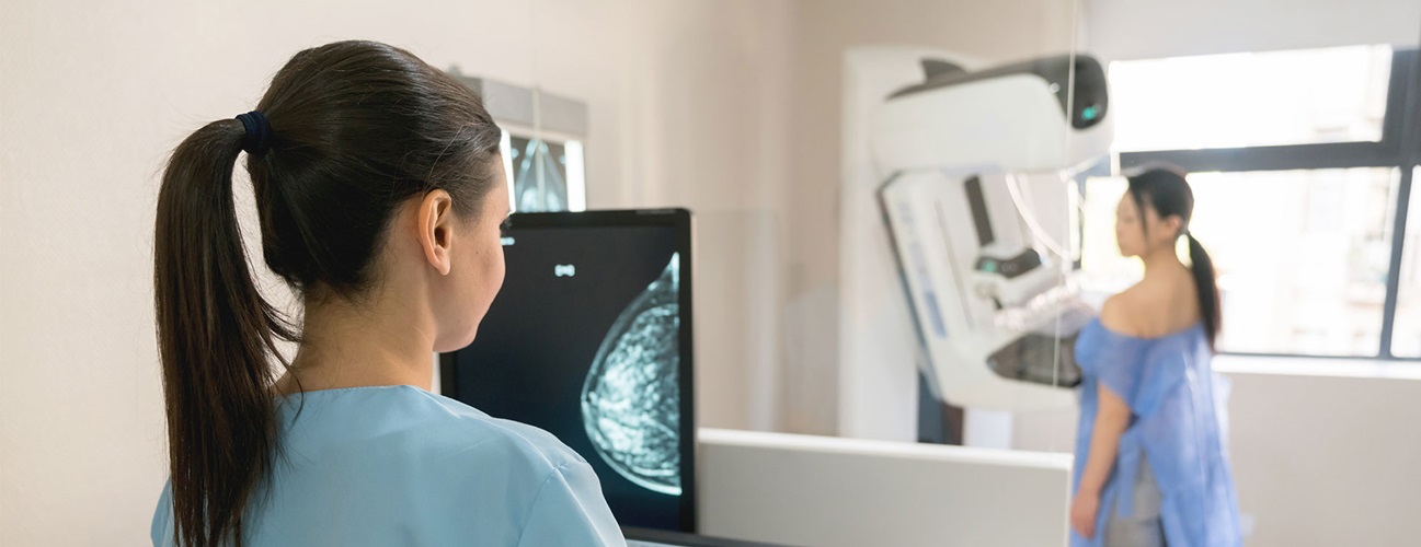
Mammography Procedure: What to Expect
Here is what happens during a typical screening mammogram:
- You will be asked to remove your clothing from the waist up and given a gown to wear, keeping the opening in the front. You will need to remove any other clothing or jewelry that may interfere with the exam.
- After entering the room where the mammogram takes place, you will be asked to remove one arm from the gown and stand in front of the mammography machine. The X-ray technologist will place one breast on the X-ray plate and may manually move the breast into the proper position.
- A separate flat plate will be brought down on top of the breast to compress it against the X-ray plate. The more compression, the more accurate the picture. You may feel some discomfort or pressure, but this part of the process should only last for a few moments.
- Once you are properly positioned, the X-ray technologist may move behind a wall to operate the computer system that takes the pictures. They can speak to and hear you from this location.
- During your mammogram, you will be asked to hold your breath while the X-ray happens. This is only for a few seconds.
- For 2D mammograms, the X-ray machine takes an image at the top and side, while during 3D tomosynthesis, the X-ray arm sweeps in a slight arc over the breast, taking multiple images.
- The X-ray technologist will tell you to resume breathing and will loosen the plate holding your breast in the machine. Then, the process is repeated for the other breast.
- When both breasts are imaged and the X-ray technologist confirms complete and clear images have been obtained, the mammogram is over: You can get dressed and go home.
The X-ray technologist cannot tell you the results of the test or describe the findings. The results must be reviewed by a radiologist.
Video All About Mammograms
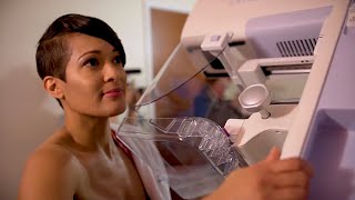
What to Expect During Your First Mammogram
A Breast Radiologist’s Perspective: Screening Mammography and Diagnostic Imaging Webinar
Let’s Talk Mammograms

What happens after my mammogram?
There is typically no special care needed after a mammogram. However, your health care provider may give you additional instructions depending on your specific health condition.
If your breasts are sore, an over-the-counter pain reliever such as acetaminophen or ibuprofen may relieve the discomfort.
With computer-aided detection (CAD) systems, a mammographic image is analyzed for masses, calcifications or areas of abnormal density that may indicate the presence of cancer. The images are highlighted by the CAD system for further analysis by the radiologist.
Getting Your Mammogram Results
The radiologist will send the report to your referring doctor. In about a week, you will get your results in a report sent to you through the mail or electronically through a patient portal. If something abnormal is found, the radiology office or your doctor will reach out to you soon after your exam to schedule a follow-up.
If a mammogram finds areas that appear abnormal, these will be noted in the report. Mammography cannot prove that an abnormal area is cancer, but further tests can be performed to narrow down the diagnosis, such as a repeat (diagnostic) mammogram, a breast ultrasound or a breast MRI.
If you are asked to come back for additional images, don’t be alarmed: A mammogram “callback” is a common occurrence. In the United States, approximately 5% to 15% of people who get mammograms are called back for additional imaging. You may be called back for more imaging after your first mammogram because the radiologists don’t have images to compare your current results to yet. The findings of this additional imaging are usually benign, meaning the changes are not caused by cancer.
If the radiologist suspects cancer after additional imaging is obtained, an ultrasound will likely be ordered to gather more information. If necessary, a bit of tissue from the suspicious area may be removed with a needle or surgery for a biopsy. A pathologist examines the tissue under a microscope to help determine if it is cancer, and if so, what type it might be.
Read more about understanding your mammogram results.
Are mammograms covered by insurance?
Most states now require that health insurance policies offer reimbursement for screening mammography. Medicare covers annual mammography screenings for women age 40 and older.
The Affordable Care Act makes mammograms for breast cancer screening available without a copay or deductible beginning with plan years starting after Aug. 1, 2012. However, health plans in place before the act was passed do not have to cover the charges for mammograms. Those plans are covered by various state and federal laws.
For those with low income, mammograms are covered through the National Breast and Cervical Cancer Early Detection Program. For more information, contact your state Department of Health. Many mammography facilities also offer special programs and lower fees during National Breast Cancer Awareness Month in October.
Who performs mammograms?
Mammograms are done by licensed, board-certified X-ray technologists. These practitioners are specially trained to capture breast images of the highest quality with the least amount of radiation exposure, while making the exam as comfortable as it can be.
X-ray technologists do not interpret the mammogram images — this part is done by a radiologist. Research shows that radiologists who specialize in breast imaging interpret mammograms with higher accuracy than those who image all body parts. You may wish to ask your facility if the radiologists specialize in breast imaging.