Lung Biopsy
What is a lung biopsy?
A biopsy is a procedure performed to remove tissue or cells from the body for examination under a microscope. A lung biopsy is a procedure in which samples of lung tissue are removed (with a special biopsy needle or during surgery) to determine if lung disease or cancer is present.
A lung biopsy may be performed using either a closed or an open method. Closed methods are performed through the skin or through the trachea (windpipe). An open biopsy is performed in the operating room under general anesthesia.
The various biopsy procedures include:
-
Needle biopsy. After a local anesthetic is given, the doctor uses a needle that is guided through the chest wall into a suspicious area with computed tomography (CT or CAT scan) or fluoroscopy (a type of X-ray “movie”) to obtain a tissue sample. This type of biopsy may also be referred to as a closed, transthoracic, or percutaneous (through the skin) biopsy.
-
Transbronchial biopsy. This type of biopsy is performed through a fiberoptic bronchoscope (a long, thin tube that has a close-focusing telescope on the end for viewing) through the main airways of the lungs (bronchoscopy).
-
Thoracoscopic biopsy. After a general anesthetic is given, an endoscope is inserted through the chest wall into the chest cavity. Various types of biopsy tools can be inserted through the endoscope to obtain lung tissue for examination. This procedure may be referred to as video-assisted thoracic surgery (VATS) biopsy. In addition to obtaining tissue for biopsy, therapeutic procedures, such as the removal of a nodule or other tissue lesion may be performed.
-
Open biopsy. After a general anesthetic is given, the doctor makes an incision in the skin on the chest and surgically removes a piece of lung tissue. Depending on the results of the biopsy, more extensive surgery, such as the removal of a lung lobe may be performed during the procedure. An open biopsy is a surgical procedure and requires a hospital stay.
Other related procedures that may be used to help diagnose problems of the lungs and respiratory tract include chest X-ray , CT scan of the chest , magnetic resonance imaging (MRI) , bronchoscopy , bronchography, chest fluoroscopy , chest ultrasound , lung scan , oximetry , mediastinoscopy , peak flow measurement , positron emission tomography (PET) scan , pulmonary function tests , pleural biopsy , pulmonary angiogram, sinus X-ray , and thoracentesis .
Expert Answers About Lung Cancer Screenings
Pulmonologist Lonny Yarmus explains who should be screened and why.
.png?h=249&iar=0&mh=360&mw=520&w=200&hash=FD12C1883B01C37077405A108F756241)
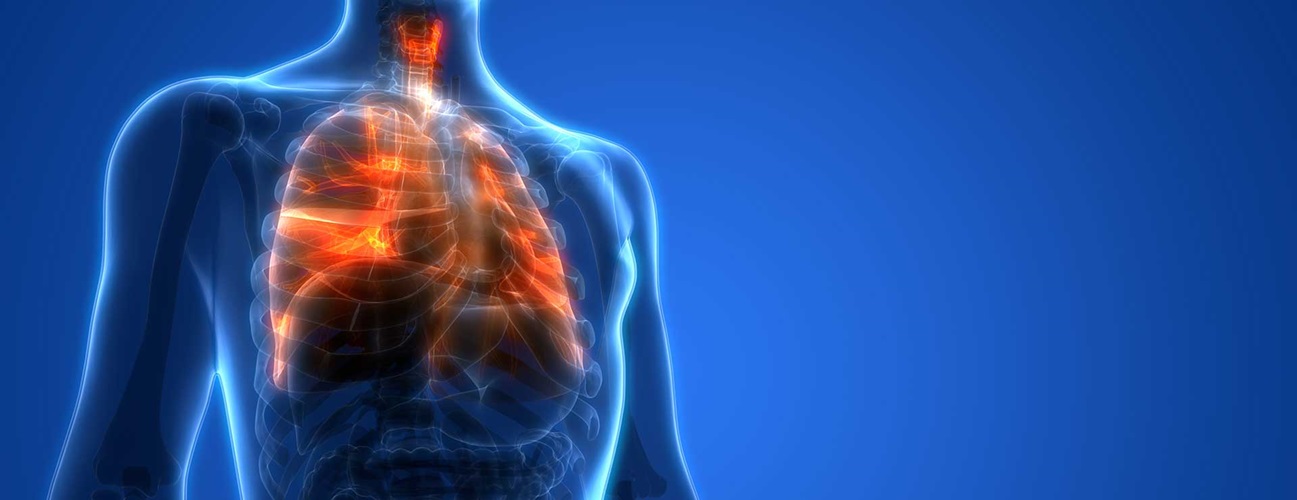
Anatomy of the respiratory system
The respiratory system is made up of the organs involved in the exchange of gases--primarily oxygen and carbon dioxide--and consists of the:
-
Nose
-
Pharynx
-
Larynx
-
Trachea
-
Bronchi
-
Lungs
The upper respiratory tract includes the:
-
Nose
-
Nasal cavity
-
Ethmoidal air cells
-
Frontal sinuses
-
Maxillary sinus
-
Larynx
-
Trachea
The lower respiratory tract includes the lungs, bronchi, and alveoli.
What are the functions of the lungs?
The lungs take in oxygen, which cells need to carry out their normal functions. The lungs also get rid of carbon dioxide, a waste product of the body's cells.
The lungs are a pair of cone-shaped organs made up of spongy, pinkish-gray tissue. They take up most of the space in the chest, or the thorax (the part of the body between the base of the neck and diaphragm).
The lungs are enveloped in a membrane called the pleura.
The lungs are separated from each other by the mediastinum, an area that contains the following:
-
The heart and its large vessels
-
Trachea (windpipe)
-
Esophagus
-
Thymus
-
Lymph nodes
The right lung has three sections called lobes. The left lung has two lobes. When you breathe, the air enters the body through the nose or the mouth. It then travels down the throat through the larynx (voice box) and trachea (windpipe) and goes into the lungs through tubes called mainstem bronchi.
One mainstem bronchus leads to the right lung and one to the left lung. In the lungs, the mainstem bronchi divide into smaller bronchi and then into even smaller tubes called bronchioles. Bronchioles end in tiny air sacs called alveoli.
Reasons for the procedure
Reasons for which a lung biopsy may be performed include, but are not limited to, the following:
-
To evaluate an abnormality seen on chest X-ray or CT scan
-
To diagnose lung infection or other lung disease
-
To investigate the cause of unexplained fluid collection in the lung
-
To determine if a lung mass is malignant (cancerous) or benign
-
To stage malignant tumors (determine the extent of spreading)
The type of biopsy performed will depend on several factors, such as the type of lung problem, the location of the lesion, and the overall condition of the person.
There may be other reasons for your doctor to recommend a lung biopsy.
Risks of the procedure
An open or thoracoscopic lung biopsy is a surgical procedure that is performed under general anesthesia. As with any surgical procedure, complications may occur. Some possible complications may include, but are not limited to, the following:
-
Blood loss or clots
-
Pain or discomfort
-
Infection
-
Pneumonia
A needle or transbronchial lung biopsy is performed under light sedation and/or local anesthesia. Some possible complications of these procedures may include, but are not limited to, the following:
-
Pneumothorax is when air becomes trapped in the pleural space causing the lung to collapse
-
Bleeding in the lung
-
Infection
If you are pregnant, or suspect that you may be pregnant, you should notify your doctor.
If the biopsy is performed using an X-ray (CT or fluoroscopy), the amount of radiation used during the procedure is considered minimal; therefore, the risk for radiation exposure is very low.
There may be other risks depending on your specific medical condition. Be sure to discuss any concerns with your doctor prior to the procedure.
Think you need a lung cancer screening?

Catching lung cancer early and treating it quickly leads to the best hope of beating the disease. Find a Johns Hopkins lung cancer expert in Baltimore and Washington, D.C., and schedule a screening today.
Before the procedure
-
The doctor will explain the procedure to you and offer you the opportunity to ask any questions that you might have.
-
You will be asked to sign a consent form that gives your permission to do the biopsy. Read the form carefully and ask questions if something is not clear. If you are to have a thoracoscopic or open lung biopsy, your doctor may discuss with you the possibility of more extensive surgery being performed during the procedure depending on the results of the biopsy.
-
In addition to a complete medical history, your doctor may perform a physical examination to ensure you are in good health before undergoing the procedure. You may undergo blood or other diagnostic tests.
-
You will be asked to fast for eight hours before the procedure, generally after midnight. If local anesthesia is to be used for the procedure, you may be permitted clear liquids in the morning of the procedure. Your doctor will give you specific instructions.
-
If you are pregnant or suspect that you may be pregnant, you should notify your doctor.
-
Notify your doctor if you are sensitive to or are allergic to any medication, latex, iodine, tape, or anesthetic agents (local and general).
-
Notify your doctor of all medications (prescription and over-the-counter) and herbal supplements that you are taking.
-
Notify your doctor if you have a history of bleeding disorders or if you are taking any anticoagulant (blood-thinning) medications, aspirin, or other medications that affect blood clotting. It may be necessary for you to stop these medications prior to the procedure.
-
You may receive a sedative prior to the procedure to help you relax. If a sedative is given, you may need someone to drive you home afterwards.
-
Based on your medical condition, your doctor may request other specific preparation.
During the procedure
A lung biopsy may be performed on an outpatient basis or as a part of your stay in a hospital. Procedures may vary depending on your condition and your doctor’s practices. In addition, some biopsies may be performed using a local anesthetic to numb the area, while others may be performed using heavy sedation or general anesthesia.
Generally, a needle lung biopsy performed through the skin follows this process:
-
You will be asked to remove any jewelry or other objects that may interfere with the procedure.
-
You will be asked to remove clothing and will be given a gown to wear.
-
An intravenous (IV) line may be started in your arm or hand.
-
You will be positioned so that the doctor can easily reach the part of the lung that will be sampled. You may be sitting up or lying down.
-
An X-ray or CT scan may be used to locate the desired biopsy site. The skin will be marked.
-
The skin over the biopsy site will be cleansed with an antiseptic solution.
-
You will feel a needle stick when the local anesthetic is injected. This may cause a brief stinging sensation.
-
You will need to hold still, avoid coughing, and hold your breath when told to during the procedure.
-
A small incision will be made over the biopsy site. The doctor will insert the biopsy needle between the ribs into the lung.
-
You may feel discomfort or pressure when the doctor enters the lung with the needle.
-
The biopsy needle will be withdrawn. Firm pressure will be applied to the biopsy site for a few minutes, until the bleeding has stopped.
-
The doctor will close the opening in the skin with sutures or adhesive strips, if necessary.
-
A sterile bandage or dressing will be applied.
-
The lung sample will be sent to the lab for examination.
-
You may have a chest X-ray taken immediately after the biopsy.
Generally, a transbronchial lung biopsy follows this process:
-
You will be asked to remove clothing and will be given a gown to wear.
-
An intravenous (IV) line may be inserted in your arm or hand.
-
Your heart rate, blood pressure, respiratory rate, and oxygen level may be monitored during the procedure.
-
You will be positioned in a sitting position or lying on your back.
-
You may receive oxygen through a nasal cannula (tube) or face mask during the procedure.
-
You may be given a sedative to make you sleepy but arousable.
-
An X-ray may be used to locate the desired biopsy site.
-
Numbing medication will be sprayed into the back of your throat to prevent gagging as the bronchoscope is passed down your trachea into the bronchi. The spray may have a bitter taste to it. Holding your breath while the doctor sprays your throat may decrease the taste.
-
You will not be able to swallow the saliva that may collect in your mouth during the procedure due to the bronchoscope in your throat. The saliva will be suctioned from your mouth from time to time.
-
The doctor will advance the bronchoscope down your throat and into the airways. As the bronchoscope is advanced, the tissues and structures will be examined.
-
You may experience some discomfort when the bronchoscope is advanced. Your airway will not be blocked.
-
One or more tissue samples will be obtained using a needle, forceps, or a brush.
-
The doctor will remove the bronchoscope.
-
The tissue sample will be sent to the lab for examination.
Generally, a thoracoscopic lung biopsy will follow this process:
-
You will be asked to remove clothing and will be given a gown to wear.
-
An intravenous (IV) line may be started in your arm or hand.
-
If there is excessive hair at the surgical site, it may be clipped off.
-
After you have been sedated, the anesthesiologist will insert a tube into your lungs so that your breathing can be assisted by a ventilator. The anesthesiologist will continuously monitor your heart rate, blood pressure, breathing, and blood oxygen level during the surgery.
-
You will be positioned on the operating table in a manner that provides the best access to the side of the chest being operated on, usually lying on the side opposite the surgical site.
-
The skin over the surgical site will be cleansed with an antiseptic solution.
-
An incision will be made in the chest for insertion of the thoracoscope. One or more additional incisions will be made at other locations on the chest for insertion of tools to be used during the procedure.
-
After the thoracoscope is inserted into the chest, the suspicious area will be located, either by the doctor’s touch (if a nodule is present) or by viewing the lung tissue through the thoracoscope.
-
Once the nodule or suspicious tissue has been located, one or more tissue samples will be taken.
-
Depending on the type of problem found or suspected, a frozen section (the tissue is quickly frozen and examined by a pathologist) may be sent to the lab for rapid examination. If the results of the frozen section indicate certain conditions, such as certain types of cancer, more extensive surgery may be performed at this time. A larger section of lung tissue or an entire lung lobe may be removed.
-
After the biopsy and any other procedures have been completed, one or more chest tubes may be inserted into the chest cavity to aid in the removal of air or fluid postoperatively.
-
The skin incision will be closed with sutures or adhesive strips.
-
A sterile bandage or dressing will be applied.
Generally, an open lung biopsy will follow this process:
-
You will be asked to remove clothing and will be given a gown to wear.
-
An intravenous (IV) line will be inserted in your arm or hand.
-
If there is excessive hair at the surgical site, it may be clipped off.
-
After you have been sedated, the anesthesiologist will insert a tube into your lungs so that your breathing can be assisted by a ventilator. The anesthesiologist will continuously monitor your heart rate, blood pressure, breathing, and blood oxygen level during the surgery.
-
A urinary catheter may be inserted into your bladder to drain urine during the procedure.
-
You will be positioned on the operating table in a manner that provides the best access to the side of the chest being operated on, usually lying on the side opposite the surgical site.
-
The skin over the surgical site will be cleansed with an antiseptic solution.
-
An incision will be made along the front of the chest at the level of the area to be examined. The incision will extend under your arm to your back.
-
When the ribs are visualized, a special instrument will be used to spread the ribs apart, leaving the lung area exposed.
-
The doctor will examine the lung. Once the suspicious area of tissue has been located, the doctor will remove a piece of tissue.
-
Depending on the type of problem found or suspected, a frozen section (the tissue is quickly frozen and examined by a pathologist) may be sent to the lab for rapid examination. If the results of the frozen section indicate certain conditions, such as certain types of cancer, more extensive surgery may be performed at this time. A larger section of lung tissue or an entire lung lobe may be removed.
-
After the biopsy and any other procedures have been completed, one or more chest tubes may be inserted into the chest cavity to aid in the removal of air or fluid postoperatively.
-
The skin incision will be closed with sutures or surgical staples.
-
A sterile bandage or dressing will be applied.
-
An epidural catheter to infuse pain medication into your back may be inserted before you leave the operating room or in the recovery room.
After the procedure
Your recovery process will vary depending on the type of procedure performed and the type of anesthesia (if any) used. If you were given general anesthesia, you will be taken to the recovery room for observation. Once your blood pressure, pulse, and breathing are stable and you are alert, you will be taken to your hospital room.
After local anesthesia or intravenous sedation, you may be discharged to your home as soon as your blood pressure, pulse, and breathing are stable. A chest X-ray may be performed immediately after the biopsy and repeated in a few hours.
If your biopsy was performed using a bronchoscope, you may have some throat discomfort. You will not be allowed to eat or drink anything until your gag reflex has returned. You may notice some soreness of your throat and pain with swallowing for a few days. This soreness is normal. Using throat lozenges or gargle may help.
After a transbronchial lung biopsy, you may be instructed to gently cough up and spit your saliva into a basin. The nurse will monitor your secretions. Your secretions may be blood tinged.
If your biopsy was performed through the skin, you may remove the bandage when instructed to do so, and bathe as usual.
The biopsy site may be tender or sore for several days after a needle biopsy. Take a pain reliever for soreness as recommended by your doctor. Aspirin or certain other pain medications may increase the chance of bleeding. Be sure to take only recommended medications.
Your doctor may ask you to avoid strenuous physical activity for a few days.
Report any of the following to your doctor:
-
Shortness of breath
-
Chest pain
-
Difficulty breathing or pain with breathing
-
Coughing up blood
-
Fever and/or chills
-
Redness, swelling, or bleeding or other drainage from the biopsy site
Your doctor may give you additional or alternate instructions after the procedure, depending on your particular situation.