Magnetic Resonance Imaging (MRI) of the Spine and Brain
What is magnetic resonance imaging (MRI)?
(MRI of the Brain, MRI of the Spine)
Magnetic resonance imaging (MRI) is a diagnostic procedure that uses a combination of a large magnet, radiofrequencies, and a computer to produce detailed images of organs and structures within the body. Unlike X-rays or computed tomography (CT scans), MRI does not use ionizing radiation. Some MRI machines look like narrow tunnels, while others are more spacious or wider. MRI scans can last from 30 minutes to two hours.
How does an MRI scan work?
The MRI machine is a large, cylindrical (tube-shaped) machine that creates a strong magnetic field around the patient. The magnetic field, along with radio waves, alters the hydrogen atoms' natural alignment in the body. Pulses of radio waves sent from a scanner knock the nuclei in your atoms out of their normal position. As the nuclei realign back into proper position, the nuclei send out radio signals. These signals are received by a computer that analyzes and converts them into a two-dimensional (2D) image of the body structure or organ being examined.
Magnetic resonance (MRI) may be used instead of computed tomography (CT) in situations where organs or soft tissue are being studied, because MRI is better at telling the difference between normal and abnormal soft tissue.
New uses and indications for MRI have contributed to the development of additional magnetic resonance technology. Magnetic resonance angiography (MRA) is a new procedure used to evaluate blood flow through arteries in a noninvasive (the skin is not pierced) manner. MRA can also be used to detect intracranial (within the brain) aneurysms and vascular malformations (abnormalities of blood vessels within the brain, spinal cord, or other parts of the body).
Magnetic resonance spectroscopy (MRS) is another noninvasive procedure used to assess chemical abnormalities in body tissues, such as the brain. MRS may be used to assess disorders such as HIV infection of the brain , stroke , head injury , coma, Alzheimer's disease , tumors, and multiple sclerosis .
Functional magnetic resonance imaging of the brain (fMRI) is used to determine the specific location of the brain where a certain function, such as speech or memory, occurs. The general areas of the brain in which such functions occur are known, but the exact location may vary from person to person. During functional resonance imaging of the brain, you will be asked to perform a specific task, such as recite the Pledge of Allegiance, while the scan is being done. By pinpointing the exact location of the functional center in the brain, doctors can plan surgery or other treatments for a particular disorder of the brain.
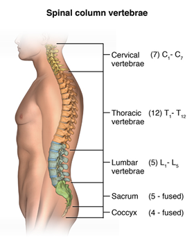
Anatomy of the spine
The spinal column, also called the vertebral or spinal canal, is made up of 33 vertebrae that are separated by spongy disks and classified into distinct areas.
-
The cervical area consists of 7 vertebrae in the neck.
-
The thoracic area consists of 12 vertebrae in the chest area.
-
The lumbar area consists of 5 vertebrae in the lower back area.
-
The sacrum has 5 small, fused vertebrae.
-
The 4 coccygeal vertebrae fuse to form 1 bone, called the coccyx or tailbone.
The spinal cord, a major part of the central nervous system, is located in the vertebral canal and reaches from the base of the skull to the upper part of the lower back. The spinal cord is surrounded by the bones of the spine and a sac containing cerebrospinal fluid. The spinal cord carries sensory and movement signals to and from the brain, and controls many reflexes.
Anatomy of the brain
The central nervous system (CNS) consists of the brain and spinal cord. The brain is an important organ that controls thought, memory, emotion, touch, motor skills, vision, respirations, temperature, hunger, and every other process that regulates our body.
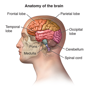
What are the different parts of the brain?
The brain can be divided into the cerebrum, brainstem, and cerebellum:
-
Cerebrum. The cerebrum (supratentorial or front of brain) is composed of the right and left hemispheres. Functions of the cerebrum include: initiation of movement, coordination of movement, body temperature, touch, vision, hearing, judgment, reasoning, problem solving, emotions, and learning.
-
Brainstem. The brainstem (midline or middle of brain) includes the midbrain, the pons, and the medulla. Functions of this area include: movement of the eyes and mouth, relaying sensory messages (such as hot, pain, and loud), hunger, respirations, consciousness, cardiac function, body temperature, involuntary muscle movements, sneezing, coughing, vomiting, and swallowing.
-
Cerebellum. The cerebellum (infratentorial or back of brain) is located at the back of the head. Its function is to coordinate voluntary muscle movements and to maintain posture, balance, and equilibrium.
More specifically, other parts of the brain include the following:
-
Pons. A deep part of the brain, located in the brainstem, the pons contains many of the control areas for eye and face movements.
-
Medulla. The lowest part of the brainstem, the medulla is the most vital part of the entire brain and contains important control centers for the heart and lungs.
-
Spinal cord. A large bundle of nerve fibers located in the back that extends from the base of the brain to the lower back, the spinal cord carries messages to and from the brain and controls many reflexes.
-
Frontal lobe. The largest section of the brain located in the front of the head, the frontal lobe is involved in personality characteristics and movement.
-
Parietal lobe. The middle part of the brain, the parietal lobe helps a person to identify objects and understand spatial relationships (where one's body is compared to objects around the person). The parietal lobe is also involved in interpreting pain and touch in the body.
-
Occipital lobe. The occipital lobe is the back part of the brain that is involved with vision.
-
Temporal lobe. The sides of the brain, these temporal lobes are involved in memory, speech, and sense of smell.
What are the reasons for an MRI of the brain or spine?
MRI may be used to examine the brain and/or spinal cord for injuries or the presence of structural abnormalities or certain other conditions, such as:
-
Tumors
-
Abscesses
-
Congenital abnormalities
-
Venous malformations
-
Hemorrhage, or bleeding into the brain or spinal cord
-
Subdural hematoma (an area of bleeding just under the dura mater, or covering of the brain)
-
Degenerative diseases, multiple sclerosis , hypoxic encephalopathy (dysfunction of the brain due to a lack of oxygen), or encephalomyelitis (inflammation or infection of the brain and/or spinal cord)
-
Hydrocephalus , or fluid in the brain
-
Herniation or degeneration of discs of the spinal cord
-
Help plan surgeries on the spine, such as decompression of a pinched nerve or spinal fusion
MRI can also help to identify the specific location of a functional center of the brain (the specific part of the brain controlling a function, such as speech or memory) to assist in treatment of a condition of the brain.
There may be other reasons for your doctor to recommend MRI of the spine or brain.
What are the risks of an MRI?
Because radiation is not used, there is no risk of exposure to ionizing radiation during an MRI exam.
Due to the use of the strong magnet, special precautions must be taken to perform an MRI on patients with certain implanted devices such as pacemakers or cochlear implants. The MRI technologist will need some information from you regarding the implanted device, such as the make and model number, to determine if it is safe for you to have an MRI. Patients who have internal metal objects, such as surgical clips, plates, screws or wire mesh, might not be eligible for an MRI.
If there is a possibility that you are claustrophobic, then you can ask your physician to provide you with anti-anxiety medication to take prior to your MRI examination. You should plan to have someone drive you home after the MRI.
If you are pregnant or suspect that you may be pregnant, you should notify your health care provider. To date there is no information indicating that MRI is harmful to an unborn child, however MRI testing during the first trimester is discouraged.
A doctor may order a contrast dye to be used during some MRI exams in order for the radiologist to better view internal tissues and blood vessels on the completed images. If contrast is used, there is a risk for allergic reaction. Patients who are allergic or sensitive to contrast dye or iodine should notify the radiologist or technologist.
There may be other risks depending on your specific medical condition. Be sure to discuss any concerns with your doctor prior to the procedure.
How do I prepare for an MRI?
EAT/DRINK : You may eat, drink and take medications as usual for most MRI exams. There are some specialty MRI exams that require certain restrictions. You will be provided detailed preparations instructions by Johns Hopkins Medical Imaging when you schedule your exam.
CLOTHING : You must completely change into a patient gown and lock up all personal belongings. A locker will be provided for you to use. Please remove all piercings and leave all jewelry and valuables at home.
WHAT TO EXPECT : Imaging takes place inside of a large tube-like structure, open on both ends. You must lie perfectly still for quality images. Due to the loud noise of the MRI machine, earplugs are required and will be provided.
ALLERGY : If you have had an allergic reaction to contrast that required medical treatment, contact your ordering physician to obtain the recommended prescription. You will likely take this by mouth 24, 12 and two hours prior to examination.
ANTI-ANXIETY MEDICATION : If you require anti-anxiety medication due to claustrophobia, contact your ordering physician for a prescription. Please note that you will need some else to drive you home.
STRONG MAGNETIC ENVIRONMENT : If you have metal within your body that was not disclosed prior to your appointment, your study may be delayed, rescheduled or cancelled upon your arrival until further information can be obtained.
Based on your medical condition, your health care provider may require other specific preparation.
When you call to make an appointment, it is extremely important that you inform if any of the following apply to you:
-
You have a pacemaker or have had heart valves replaced
-
You have any type of implantable pump, such as an insulin pump
-
You have vessel coils, filters, stents, or clips
-
You are pregnant or think you might be pregnant
-
You have any body piercing
-
You are wearing a medication patch
-
You have permanent eye liner or tattoos
-
You have ever had a bullet wound
-
You have ever worked with metal (for example, a metal grinder or welder)
-
You have metallic fragments anywhere in the body
-
You are not able to lie down for 30 to 60 minutes.
.png?h=231&iar=0&mh=360&mw=520&w=350&hash=0638B0A89179018E965F38A10B7F9328)
What happens during an MRI?
MRI may be performed on an outpatient basis or as part of your stay in a hospital. Procedures may vary depending on your condition and your doctor's practices.
Generally, MRI follows this process:
-
You will be asked to remove any clothing, jewelry, eyeglasses, hearing aids, hairpins, removable dental work, or other objects that may interfere with the procedure.
-
If you are asked to remove clothing, you will be given a gown to wear.
-
If you are to have a procedure done with contrast, an intravenous (IV) line will be started in the hand or arm for injection of the contrast dye.
-
You will lie on a scan table that slides into a large circular opening of the scanning machine. Pillows and straps may be used to prevent movement during the procedure.
-
The technologist will be in another room where the scanner controls are located. However, you will be in constant sight of the technologist through a window. Speakers inside the scanner will enable the technologist to communicate with and hear you. You will have a call button so that you can let the technologist know if you have any problems during the procedure. The technologist will be watching you at all times and will be in constant communication.
-
You will be given earplugs or a headset to wear to help block out the noise from the scanner. Some headsets may provide music for you to listen to.
-
During the scanning process, a clicking noise will sound as the magnetic field is created and pulses of radio waves are sent from the scanner.
-
It will be important for you to remain very still during the examination, as any movement could cause distortion and affect the quality of the scan.
-
At intervals, you may be instructed to hold your breath, or to not breathe, for a few seconds, depending on the body part being examined. You will then be told when you can breathe. You should not have to hold your breath for longer than a few seconds.
-
If contrast dye is used for your procedure, you may feel some effects when the dye is injected into the IV line. These effects include a flushing sensation or a feeling of coldness, a salty or metallic taste in the mouth, a brief headache, itching, or nausea and/or vomiting. These effects usually last for a few moments.
-
You should notify the technologist if you feel any breathing difficulties, sweating, numbness, or heart palpitations.
-
Once the scan is complete, the table will slide out of the scanner and you will be assisted off the table.
-
If an IV line was inserted for contrast administration, the line will be removed.
While the MRI procedure itself causes no pain, having to lie still for the length of the procedure might cause some discomfort or pain, particularly in the case of a recent injury or invasive procedure such as surgery. The technologist will use all possible comfort measures and complete the procedure as quickly as possible to minimize any discomfort or pain.
What happens after an MRI?
You should move slowly when getting up from the scanner table to avoid any dizziness or lightheadedness from lying flat for the length of the procedure.
If any sedatives were taken for the procedure, you may be required to rest until the sedatives have worn off. You will also need to avoid driving.
If contrast dye is used during your procedure, you may be monitored for a period of time for any side effects or reactions to the contrast dye, such as itching, swelling, rash, or difficulty breathing.
If you notice any pain, redness, and/or swelling at the IV site after you return home following your procedure, you should notify your doctor as this could indicate an infection or other type of reaction.
Otherwise, there is no special type of care required after a MRI scan of the spine and brain. You may resume your usual diet and activities, unless your doctor advises you differently.
Your doctor may give you additional or alternate instructions after the procedure, depending on your particular situation.