Pelvic Ultrasound
What is a pelvic ultrasound?
(Ultrasound-Pelvis, Pelvic Ultrasonography, Pelvic Sonography, Pelvic Scan, Lower Abdomen Ultrasound, Gynecologic Ultrasound, Transabdominal Ultrasound, Transvaginal Ultrasound, Endovaginal Ultrasound)
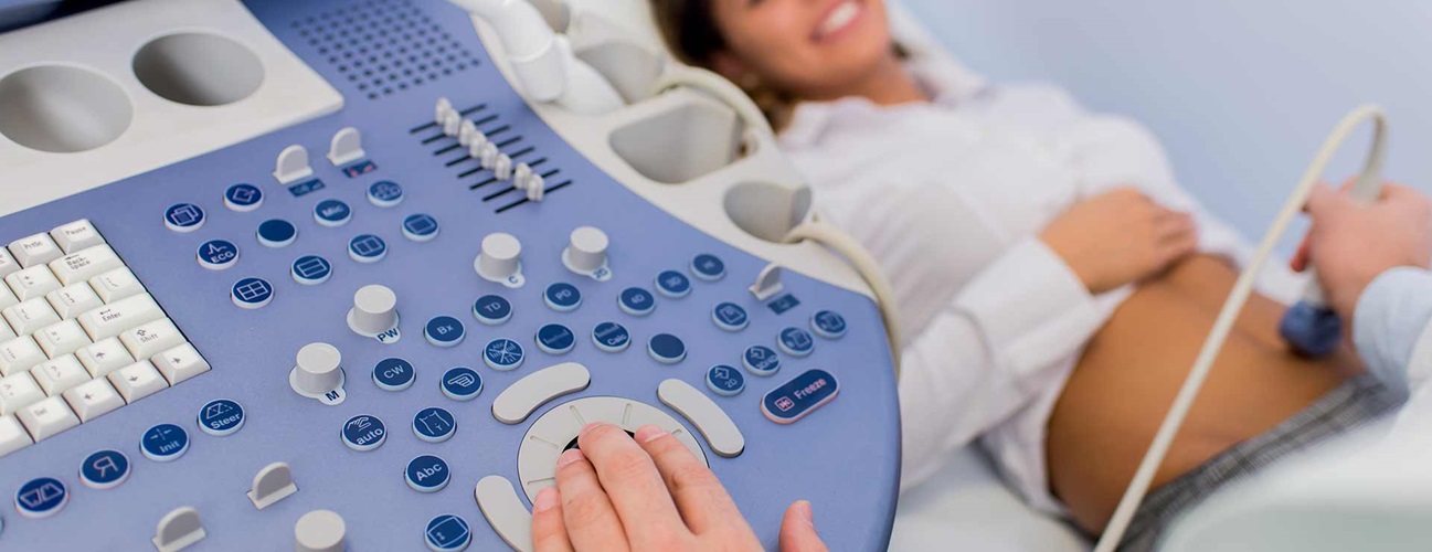
A pelvic ultrasound is a noninvasive diagnostic exam that produces images that are used to assess organs and structures within the female pelvis. A pelvic ultrasound allows quick visualization of the female pelvic organs and structures including the uterus, cervix, vagina, fallopian tubes and ovaries.
Ultrasound uses a transducer that sends out ultrasound waves at a frequency too high to be heard. The ultrasound transducer is placed on the skin, and the ultrasound waves move through the body to the organs and structures within. The sound waves bounce off the organs like an echo and return to the transducer. The transducer processes the reflected waves, which are then converted by a computer into an image of the organs or tissues being examined.
The sound waves travel at different speeds depending on the type of tissue encountered - fastest through bone tissue and slowest through air. The speed at which the sound waves are returned to the transducer, as well as how much of the sound wave returns, is translated by the transducer as different types of tissue.
An ultrasound gel is placed on the transducer and the skin to allow for smooth movement of the transducer over the skin and to eliminate air between the skin and the transducer for the best sound conduction.
Another type of ultrasound is Doppler ultrasound, sometimes called a duplex study, used to show the speed and direction of blood flow in certain pelvic organs. Unlike a standard ultrasound, some sound waves during the Doppler exam are audible.
Pelvic ultrasound may be performed using one or both of 2 methods:
-
Transabdominal (through the abdomen). A transducer is placed on the abdomen using the conductive gel
-
Transvaginal (through the vagina). A long, thin transducer is covered with the conducting gel and a plastic/latex sheath and is inserted into the vagina
The type of ultrasound procedure performed depends on the reason for the ultrasound. Only one method may be used, or both methods may be needed to provide the information needed for diagnosis or treatment.
Other related procedures that may be used to evaluate problems of the pelvis include hysteroscopy , colposcopy , and laparoscopy .
.png?h=200&iar=0&mh=360&mw=520&w=200&hash=E8C7D43634560907435FD3759F98A7FF)
What are female pelvic organs?
The organs and structures of the female pelvis are:
-
Endometrium. The lining of the uterus
-
Uterus (also known as the womb). The uterus is a hollow, pear-shaped organ located in a woman's lower abdomen, between the bladder and the rectum. It sheds its lining each month during menstruation, unless a fertilized egg (ovum) becomes implanted and pregnancy follows.
-
Ovaries. Two female reproductive organs located in the pelvis in which egg cells (ova) develop and are stored and where the female sex hormones estrogen and progesterone are produced.
-
Cervix. The lower, narrow part of the uterus located between the bladder and the rectum, forming a canal that opens into the vagina, which leads to the outside of the body.
-
Vagina (also known as the birth canal). The passageway through which fluid passes out of the body during menstrual periods. The vagina connects the cervix and the vulva (the external genitalia).
-
Vulva. The external portion of the female genital organs
What are the reasons for a pelvic ultrasound?
Pelvic ultrasound may be used for measurement and evaluation of female pelvic organs. Ultrasound assessment of the pelvis may include, but is not limited to, the following:
-
Size, shape, and position of the uterus and ovaries
-
Thickness, echogenicity (darkness or lightness of the image related to the density of the tissue), and presence of fluids or masses in the endometrium, myometrium (uterine muscle tissue), fallopian tubes, or in or near the bladder
-
Length and thickness of the cervix
-
Changes in bladder shape
-
Blood flow through pelvic organs
Pelvic ultrasound can provide much information about the size, location, and structure of pelvic masses, but cannot provide a definite diagnosis of cancer or specific disease. A pelvic ultrasound may be used to diagnose and assist in the treatment of the following conditions:
-
Abnormalities in the anatomic structure of the uterus, including endometrial conditions
-
Fibroid tumors (benign growths), masses, cysts, and other types of tumors within the pelvis
-
Presence and position of an intrauterine contraceptive device (IUD)
-
Pelvic inflammatory disease (PID) and other types of inflammation or infection
-
Postmenopausal bleeding
-
Monitoring of ovarian follicle size for infertility evaluation
-
Aspiration of follicle fluid and eggs from ovaries for in vitro fertilization
-
Ectopic pregnancy (pregnancy occurring outside of the uterus, usually in the fallopian tube)
-
Monitoring fetal development during pregnancy
-
Assessing certain fetal conditions
Ultrasound may also be used to assist with other procedures such as endometrial biopsy . Transvaginal ultrasound may be used with sonohysterography, a procedure in which the uterus is filled with fluid to distend it for better imaging.
There may be other reasons for your doctor to recommend a pelvic ultrasound.
What are the risks of a pelvic ultrasound?
There is no radiation used and generally no discomfort from the application of the ultrasound transducer to the skin during a transabdominal ultrasound. You may experience slight discomfort with the insertion of the transvaginal transducer into the vagina.
Transvaginal ultrasound requires covering the ultrasound transducer in a plastic or latex sheath, which may cause a reaction in patients with a latex allergy.
During a transabdominal ultrasound, you may experience discomfort from having a full bladder or lying on the examination table.
If a transabdominal ultrasound is needed quickly, a urinary catheter may be inserted to fill the bladder.
There may be risks depending on your specific medical condition. Be sure to discuss any concerns with your doctor prior to the procedure.
Certain factors or conditions may interfere with the results of the test. These include, but are not limited to, the following:
-
Severe obesity
-
Barium within the intestines from a recent barium procedure
-
Inadequate filling of bladder (with transabdominal ultrasound). A full bladder helps move the uterus up and moves the bowel away for better imaging.
How do I prepare for a pelvic ultrasound?
EAT/DRINK : Drink a minimum of 24 ounces of clear fluid at least one hour before your appointment. Do not empty your bladder until after the exam.
Generally, no fasting or sedation is required for a pelvic ultrasound, unless the ultrasound is part of another procedure that requires anesthesia.
For a transvaginal ultrasound, you should empty your bladder right before the procedure.
Your doctor will explain the procedure to you and offer you the opportunity to ask any questions that you might have about the procedure.
Based on your medical condition, your doctor may request other specific preparation.
What happens during a pelvic ultrasound?
A pelvic ultrasound may be performed in your doctor’s office, on an outpatient basis, or as part of your stay in a hospital. Procedures may vary depending on your condition and your hospital’s practices.
Generally, a pelvic ultrasound follows this process:
/getimage-(9).png?h=200&iar=0&mh=360&mw=520&w=200&hash=94913919204845A71B0269C945C366F5)
For a transabdominal ultrasound
-
You will be asked to remove any clothing, jewelry, or other objects that may interfere with the scan.
-
If asked to remove clothing, you will be given a gown to wear.
-
You will lie on your back on an examination table.
-
A gel-like substance will be applied to your abdomen.
-
The transducer will be pressed against the skin and moved around over the area being studied.
-
If blood flow is being assessed, you may hear a "whoosh, whoosh" sound when the Doppler probe is used.
-
Images of structures will be displayed on the computer screen. Images will be recorded on various media for the health care record.
-
Once the procedure has been completed, the gel will be removed.
-
You may empty your bladder when the procedure is completed.
.png?h=200&iar=0&mh=360&mw=520&w=200&hash=3CAB3596EC446D68FE4C819E7C2B9AAA)
For a transvaginal ultrasound
-
You will be asked to remove any clothing, jewelry, or other objects that may interfere with the scan.
-
If asked to remove clothing, you will be given a gown to wear.
-
You will lie on an examination table, with your feet and legs supported as for a pelvic examination.
-
A long, thin transvaginal transducer will be covered with a plastic or latex sheath and lubricated. The tip of the transducer will be inserted into your vagina. This may be slightly uncomfortable.
-
The transducer will be gently turned and angled to bring the areas for study into focus. You may feel mild pressure as the transducer is moved.
-
If blood flow is being assessed, you may hear a "whoosh, whoosh" sound when the Doppler probe is used.
-
Images of organs and structures will be displayed on the computer screen. Images may be recorded on various media for the health care record.
-
Once the procedure has been completed, the transducer will be removed.
What happens after a pelvic ultrasound?
There is no special type of care required after a pelvic ultrasound. You may resume your normal diet and activity unless your doctor advises you differently.
There are no confirmed adverse biological effects on patients or instrument operators caused by exposures to ultrasound at the intensity levels used in a diagnostic ultrasound.
Your doctor may give you additional or alternate instructions after the procedure, depending on your particular situation.