Magnetic Resonance Imaging (MRI) of the Heart
What is MRI of the heart?
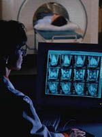
Magnetic resonance imaging (MRI) is a test that uses a large magnet, radio signals, and a computer to make images of organs and tissue in the body. In this case, the heart is imaged.
The MRI machine is large and tube-shaped. It creates a strong magnetic field around the body. Some MRI machines are more open.
The magnetic field lines up the hydrogen protons in your body. The radio waves then knock the protons out of position. As they realign back into proper position, they send out radio signals. A computer receives the signals and converts them into images of the body. This image appears on a viewing monitor.
MRI may be used instead of a CT scan when organs or soft tissues are being studied.
Why might I need an MRI of the heart?
MRI of the heart may be done to assess signs or symptoms that may suggest:
-
Atherosclerosis. This is a gradual clogging of the arteries by fatty materials and other substances in the blood stream. It develops over many years.
-
Cardiomyopathy. This happens when the heart muscle becomes thick and weakened.
-
Congenital heart disease. These are defects in the heart that happen as the fetus forms. An example is a hole in the wall between the two lower chambers of the heart (ventricular septal defect).
-
Heart failure. This condition means the heart muscle is weak and can’t pump enough blood to the body.
-
Aneurysm. This is a widening and weakening of a part of the heart muscle or the aorta.
-
Heart valve disease. When heart valves become damaged, it can block blood flow in the heart.
-
Cardiac tumor. A tumor of the heart may happen on the outside surface or inside the heart.
There may be other reasons for your healthcare provider to recommend an MRI of the heart.
#TomorrowsDiscoveries: Cardiovascular Imaging | Joao A. C. Lima, M.D., M.B.A.

Dr. Joao Lima describes recent advances in imaging that provide crucial information about the heart and blood vessels without the need for invasive procedures like dye injections and catheterization.
What are the risks of a MRI of the heart?
There is no radiation exposure during MRI.
You can’t have an MRI if you have a:
-
Older intracranial aneurysm clips
-
Cochlear implants
-
Certain prosthetic devices (such as artificial joint)
-
Implanted medicine infusion pump
-
Neurostimulator
-
Bone growth stimulator
-
Certain intrauterine contraceptives (IUDs)
-
Other iron-based metal implants
-
Bullet or shrapnel
Patients with implanted cardiac pacemakers and defibrillators can undergo an MRI but will need special considerations based on the type of device the patient has and the MRI equipment. It is advised your referring physician contacts the MRI technologist or radiologist.
If you are pregnant or think you may be, tell your healthcare provider. MRI is generally safe in pregnancy, but you and your healthcare provider should discuss the risks and benefits of having MRI.
If contrast dye is used, there is a risk you could have an allergic reaction to the dye. If you are allergic to or sensitive to medicines, tell your healthcare provider. If you have kidney problems, there is a risk of a serious reaction to the dye. Discuss this risk with your healthcare provider prior to the test.
MRI contrast may have an effect on other conditions, such as allergies, asthma, anemia, low blood pressure, kidney disease, and sickle cell disease.
Nephrogenic systemic fibrosis (NSF) is a very rare but serious complication of MRI contrast use in people with kidney disease or kidney failure. If you have a history of kidney disease, kidney failure, kidney transplant, liver disease or are on dialysis, inform the MRI technologist or radiologist prior to receiving contrast.
There may be other risks depending on your specific medical condition. Be sure to discuss any concerns with your healthcare provider prior to the MRI.
How do I prepare for an MRI?
EAT/DRINK: You may eat, drink and take medications as usual for most MRI exams. There are some specialty MRI exams that require certain restrictions. You will be provided detailed preparations instructions by Johns Hopkins Medical Imaging when you schedule your exam.
CLOTHING: You must completely change into a patient gown and lock up all personal belongings. A locker will be provided for you to use. Please remove all piercings and leave all jewelry and valuables at home.
WHAT TO EXPECT: Imaging takes place inside of a large tube-like structure, open on both ends. You must lie perfectly still for quality images. Due to the loud noise of the MRI machine, earplugs are required and will be provided.
ALLERGY: If you have had an allergic reaction to contrast that required medical treatment, contact your ordering physician to obtain the recommended prescription. You will likely take this by mouth 24, 12 and two hours prior to examination.
ANTI-ANXIETY MEDICATION: If you require anti-anxiety medication due to claustrophobia, contact your ordering physician for a prescription. Please note that you will need some else to drive you home.
STRONG MAGNETIC ENVIRONMENT: If you have metal within your body that was not disclosed prior to your appointment, your study may be delayed, rescheduled or cancelled upon your arrival until further information can be obtained.
Based on your medical condition, your health care provider may require other specific preparation.
When you call to make an appointment, it is extremely important that you inform if any of the following apply to you:
-
You have a pacemaker or have had heart valves replaced
-
You have any type of implantable pump, such as an insulin pump
-
You have vessel coils, filters, stents, or clips
-
You are pregnant or think you might be pregnant
-
You have any body piercing
-
You are wearing a medication patch
-
You have permanent eye liner or tattoos
-
You have ever had a bullet wound
-
You have ever worked with metal (for example, a metal grinder or welder)
-
You have metallic fragments anywhere in the body
-
You are not able to lie down for 30 to 60 minutes.
What happens during an MRI of the heart?
You may have your MRI an outpatient basis or as part of your stay in a hospital.
Generally, an MRI follows this process:
-
Remove any clothing, jewelry, eyeglasses, hearing aids, hairpins, removable dental work, or other objects that may interfere with the MRI.
-
If you are asked to remove clothing, you will be given a gown to wear.
-
If you are to have a MRI done with contrast, a nurse will start an intravenous (IV) line in the hand or arm to inject the contrast dye.
-
You will lie on a scan table that slides into a large circular opening of the scanning machine. You may have pillows or straps to prevent movement during the MRI.
-
The technologist will be in another room where the scanner controls are located. However, you will be in constant sight of the technologist through a window. Speakers inside the scanner will enable the technologist to talk to and hear you. You will have a call button so that you can let the technologist know if you have any problems during the MRI. The technologist will be watching you at all times and will be in constant communication.
-
You will be given earplugs or a headset to wear to help block out the noise from the scanner. You may be able to listen to music.
-
During the scanning process, a clicking noise will sound as the magnetic field is created and pulses of radio waves are sent from the scanner.
-
It’s important to remain very still during the test. Any movement could affect the quality of the scan.
-
You may be told to hold your breath, or to not breathe, for a few seconds. You will then be told when you can breathe. You should not have to hold your breath for longer than a few seconds.
-
If contrast dye is used, you may feel some effects when the dye is injected into the IV line. These include coolness or discomfort at the IV site, and should only last for a few moments.
-
You should tell the technologist if you feel any breathing difficulties, sweating, numbness, or heart palpitations.
-
Once the scan is done, the table will slide out of the scanner and you will be helped off the table.
-
If an IV line was inserted to give contrast dye, the line will be removed.
The MRI itself causes no pain. But, having to lie still for might be uncomfortable. The technologist will use all possible comfort measures and complete the test as quickly as possible to reduce any discomfort or pain.
If you have metal fillings in your teeth, you may feel some slight tingling of the teeth during the test.
What happens after an MRI of the heart?
Move slowly when getting up from the scanner table to avoid any dizziness or lightheadedness from lying flat for the length of the MRI.
If you had a sedative, you need to rest until the sedatives have worn off. You will also need to avoid driving.
If contrast dye is used, you may be watched for a period for any side effects or reactions to the contrast dye, such as itching, swelling, rash, or difficulty breathing.
If you notice any pain, redness, and/or swelling at the IV site after you return home, tell your healthcare provider. It could mean you have an infection or other type of reaction.
Otherwise, there is no special type of care required after a MRI scan of the heart. You may go back your usual diet and activities, unless your healthcare provider advises you differently.
Your healthcare provider may give you other instructions after the MRI, depending on your situation.