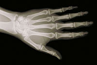
Chest X-Ray
What is a chest X-ray?
A chest X-ray is an imaging test that uses X-rays to look at the structures and organs in your chest. It can help your healthcare provider see how well your lungs and heart are working. Certain heart problems can cause changes in your lungs. Certain diseases can cause changes in the structure of the heart or lungs.
Chest X-rays can show your healthcare provider the size, shape, and location of the following:
-
Heart
-
Lungs
-
Bronchi
-
Aorta
-
Pulmonary arteries
-
Middle chest area (mediastinum)
-
Bones of your chest
It uses a small amount of radiation to make pictures of these areas.
Why might I need a chest X-ray?
Your healthcare provider may order a chest X-ray to see how well your heart or lungs are working. You may need a chest X-ray if it is suspected that you have any of the following:
-
Enlarged heart which can mean you have a congenital heart defect or cardiomyopathy
-
Fluid in the space between your lungs and your chest wall (pleural effusion)
-
Pneumonia or another lung problem
-
Ballooning of the aorta or another great blood vessel (aneurysm)
-
Broken bone
-
Hardening of a heart valve or aorta (calcification)
-
Tumors or cancer
-
Diaphragm that has moved out of place (hernia)
-
Inflammation of the lining of the lung (pleuritis)
-
Fluid in the lungs (pulmonary edema) which can mean you have congestive heart failure
You may also need a chest X-ray:
-
As part of a complete physical exam or before you have surgery
-
To check on symptoms related to the heart or lungs
-
To see how well treatment if working or how a disease is progressing
-
To check on your lungs and chest cavity after surgery
-
To see where implanted pacemaker wires and other internal devices are located
These other devices include central venous catheters, endotracheal tubes, chest tubes, and nasogastric tubes.
Your healthcare provider may have other reasons to recommend a chest X-ray.
What are the risks of a chest X-ray?
You may want to ask your healthcare provider about the amount of radiation used during the test. Also ask about the risks as they apply to you.
Consider writing down all X-rays you get, including past scans and X-rays for other health reasons. Show this list to your healthcare provider. The risks of radiation exposure may be tied to the number of X-rays you have and the X-ray treatments you have over time.
Tell your healthcare provider if you are pregnant or think you may be pregnant. Radiation exposure during pregnancy may lead to birth defects.
You may have other risks depending on your specific health condition. Talk with your healthcare provider about any concerns you have before the procedure.
How do I get ready for a chest X-ray?
-
Your healthcare provider will explain the procedure to you. Ask any questions you have about the procedure.
-
You may be asked to sign a consent form that gives permission to do the procedure. Read the form carefully and ask questions if anything is not clear.
-
You usually do not need to stop eating or drinking before the test. You also usually will not need medicine to help you relax (sedation).
-
Tell your healthcare provider if you are pregnant or think you may be pregnant.
-
Wear clothing that you can easily take off. Or wear clothing that lets the radiologist reach your chest.
-
Tell your healthcare provider if you have any body piercings on your chest.
-
Follow any other instructions your healthcare provider gives you to get ready.
What happens during a chest X-ray?
You may have a chest X-ray as an outpatient or as part of your stay in a hospital. The way the test is done may vary depending on your condition and your healthcare provider’s practices.
Generally, a chest X-ray follows this process:
-
You will be asked to remove any clothing, jewelry, or other objects that may get in the way of the test.
-
You will be given a gown to wear.
-
You may be asked to lie down, sit, or stand. Your position depends on what images the technologist needs.
-
For a standing or sitting image, you will stand or sit in front of the X-ray plate. You will be asked to roll your shoulders forward, take in a deep breath, and hold it until the X-ray is made. If you are unable to hold your breath, the technologist will take the picture by watching how you breathe.
-
You will need to stay still during the X-ray. Moving during the X-ray may affect the quality of the image.
-
For a side-angle view of the chest, you will be asked to turn to your side and raise your arms above your head. You will be told to take in a deep breath and hold it as the X-ray is made.
-
The technologist will step behind a special window while the images are being made.
The chest X-ray is not painful. But you may have some discomfort or pain from moving into different positions if you have had recent surgery or an injury. The technologist will use all possible comfort measures and do the scan as quickly as possible to minimize any discomfort or pain.
What happens after a chest X-ray?
You do not need any special care after a chest X-ray. Your healthcare provider may give you other instructions, depending on your situation.