Lithotripsy
What is lithotripsy?
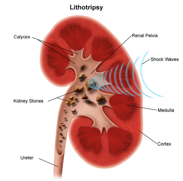
Lithotripsy is a noninvasive (the skin is not pierced) procedure used to treat kidney stones that are too large to pass through the urinary tract. Lithotripsy treats kidney stones by sending focused ultrasonic energy or shock waves directly to the stone first located with fluoroscopy (a type of X-ray “movie”) or pelvic ultrasound (high frequency sound waves). The shock waves break a large stone into smaller stones that will pass through the urinary system. Lithotripsy allows persons with certain types of stones in the urinary system to avoid an invasive surgical procedure for stone removal. In order to aim the waves, your doctor must be able to see the stones under X-ray or kidney ultrasound.
The introduction of lithotripsy in the early 1980s revolutionized the treatment of patients with kidney stone disease. Patients who once required major surgery to remove their stones could be treated with lithotripsy, and not even require an incision. As such, lithotripsy is the only non-invasive treatment for kidney stones, meaning no incision or internal telescopic device is required.
Lithotripsy involves the administration of a series of shock waves to the targeted stone. The shock waves, which are generated by a machine called a lithotripter, are focused by x-ray onto the kidney stone. The shock waves travel into the body, through skin and tissue, reaching the stone where they break it into small fragments. For several weeks following treatment, those small fragments are passed out of the body in the urine.
In the two-plus decades since lithotripsy was first performed in the United States, we have learned a great deal about how different patients respond to this technology. It turns out that we can identify some patients who will be unlikely to experience a successful outcome following lithotripsy, whereas we may predict that other patients will be more likely to clear their stones. Although many of these parameters are beyond anyone's control, such as the stone size and location in the kidney, there are other maneuvers that can be done during lithotripsy treatment that may positively influence the outcome of the procedure. At the Brady Urological Institute, our surgeons have researched techniques to make lithotripsy safer and more effective, and we incorporate our own findings as well as those of other leading groups to provide a truly state of the art treatment.
Other procedures that may be used to treat kidney stones include:
- Urethroscopy or ureteroscopy. Endoscopic procedures in which stones in the urethra or ureter may be removed with a device inserted through a short, flexible, lighted tube, called an endoscope.
- Percutaneous nephrolithotomy (tunnel surgery). A surgical procedure for stones that cannot be treated with lithotripsy or endoscopic procedures. It involves the removal of a stone through a thin tube tunneled through a small incision in the back into the kidney.
- Open surgery. A more invasive surgical procedure using a larger incision to directly access the stone.
- Stent. A synthetic, tubular device that may be used along with other procedures. A stent may be inserted through a special scope into the urinary tract to allow stones to pass more easily.
About kidney stones
When substances that are normally excreted through the kidneys remain in the urinary tract, they may crystallize and harden into a kidney stone. If the stones break free of the kidney, they can travel through, and get lodged in, the narrower passages of the urinary tract. Some kidney stones are small or smooth enough to pass easily through the urinary tract without discomfort. Other stones may have rough edges or grow as large as a pea causing extreme pain as they travel through or become lodged in the urinary tract. The areas most prone to trapping kidney stones are the bladder, ureters, and urethra.
Most kidney stones that develop are small enough to pass without intervention. However, in about 20 percent of cases, the stone is greater than 2 centimeters (about one inch) and may require treatment. Most kidney stones are composed of calcium; however, there are other types of kidney stones that can develop. Types of kidney stones include:
- Calcium stones. Calcium, a normal part of a healthy diet used in bones and muscles, is normally flushed out with the rest of the urine. However, excess calcium not used by the body may combine with other waste products to form a stone.
- Struvite stones. Struvite stones, composed of magnesium, phosphate, and ammonia, may occur after a urinary tract infection.
- Uric acid stones. Uric acid stones may occur when urine is too acidic, as in certain conditions, such as gout or malignancies.
- Cystine stones. Cystine stones consist of cystine, one of the building blocks that make up muscles, nerves, and other parts of the body.
How does the urinary system work?
The body takes nutrients from food and converts them to energy. After the body has taken the food that it needs, waste products are left behind in the bowel and in the blood.
The urinary system keeps chemicals, such as potassium and sodium, and water in balance, and removes a type of waste, called urea, from the blood. Urea is produced when foods containing protein, such as meat, poultry, and certain vegetables, are broken down in the body. Urea is carried in the bloodstream to the kidneys.
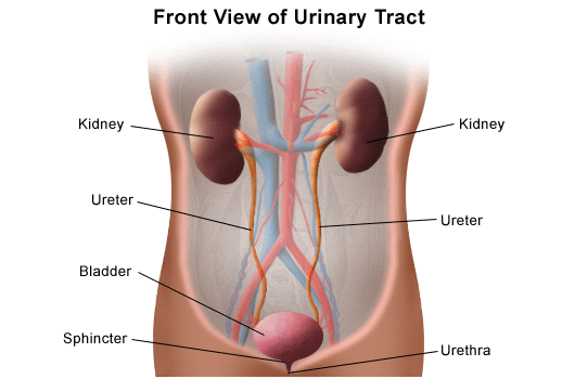
Urinary system parts and their functions:
- Two kidneys. A pair of purplish-brown organs located below the ribs toward the middle of the back. Their function is to:
- Remove liquid waste from the blood in the form of urine
- Keep a stable balance of salts and other substances in the blood
- Produce erythropoietin, a hormone that aids the formation of red blood cells
- Regulate blood pressure
The kidneys remove urea from the blood through tiny filtering units called nephrons. Each nephron consists of a ball formed of small blood capillaries, called a glomerulus, and a small tube called a renal tubule. Urea, together with water and other waste substances, forms the urine as it passes through the nephrons and down the renal tubules of the kidney.
- Two ureters. Narrow tubes that carry urine from the kidneys to the bladder. Muscles in the ureter walls continually tighten and relax forcing urine downward, away from the kidneys. If urine backs up, or is allowed to stand still, a kidney infection can develop. About every 10 to 15 seconds, small amounts of urine are emptied into the bladder from the ureters.
- Bladder. A triangle-shaped, hollow organ located in the lower abdomen. It is held in place by ligaments that are attached to other organs and the pelvic bones. The bladder's walls relax and expand to store urine, and contract and flatten to empty urine through the urethra. The typical healthy adult bladder can store up to two cups of urine for two to five hours.
- Two sphincter muscles. Circular muscles that help keep urine from leaking by closing tightly like a rubber band around the opening of the bladder
- Nerves in the bladder. These alert a person when it is time to urinate, or empty the bladder
- Urethra. The tube that allows urine to pass outside the body. The brain signals the bladder muscles to tighten, which squeezes urine out of the bladder. At the same time, the brain signals the sphincter muscles to relax to let urine exit the bladder through the urethra. When all the signals occur in the correct order, normal urination occurs.
Reasons for the procedure
The primary advantage of lithotripsy is that it is completely non-invasive.
Lithotripsy is well suited to patients with small kidney stones that can be easily seen by x-ray.
When kidney stones become too large to pass through the urinary tract, they may cause severe pain and may also block the flow of urine. An infection may develop. Lithotripsy may be performed to treat certain types of kidney stones in certain locations within the urinary tract.
There may be other reasons for your doctor to recommend lithotripsy.
Risks of the procedure
You may want to ask your doctor about the amount of radiation used during the procedure and the risks related to your particular situation. It is a good idea to keep a record of your past history of radiation exposure, such as previous scans and other types of X-rays, so that you can inform your doctor. Risks associated with radiation exposure may be related to the cumulative number of X-ray examinations and/or treatments over a long period of time.
Complications of lithotripsy may include, but are not limited to, the following:
- Bleeding around the kidney
- Infection
- Obstruction of the urinary tract by stone fragments
- Stone fragments left that may require more lithotripsies
Contraindications for lithotripsy include, but are not limited to, the following:
- Pregnant patients
- Patients on "blood thinners" or patients with bleeding disorders. Aspirin or other blood thinners must be discontinued for at least 1 week prior to lithotripsy.
- Patients with chronic kidney infection, as some fragments may not pass, so the bacteria will not be completely eliminated from the kidney.
- Patients with obstruction or scar tissue in the ureter, which may prevent stone fragments from passing.
- Patients who require immediate and/or complete clearance of stone material.
- Patients with stones composed of cystine and certain types of calcium, as these stones do not fragment well with lithotripsy
Patients with cardiac pacemakers should notify their doctor. Lithotripsy may be performed on patients with pacemakers with the approval of a cardiologist and using certain precautions. Rate-responsive pacemakers that are implanted in the abdomen may be damaged during lithotripsy.
There may be other risks depending on your specific medical condition. Be sure to discuss any concerns with your doctor prior to the procedure.
Obesity and intestinal gas may interfere with a lithotripsy procedure.
Before the procedure
- Your doctor will explain the procedure to you and offer you the opportunity to ask any questions that you might have about the procedure.
- You will be asked to sign a consent form that gives your permission to do the procedure. Read the form carefully and ask questions if something is not clear.
- In addition to a complete medical history, your doctor may perform a complete physical examination to ensure you are in good health before undergoing the procedure. You may undergo blood tests or other diagnostic tests.
- Fasting before the procedure may be indicated, depending on the type of anesthetic or sedation used. You will be given instructions on how many hours to fast before the procedure if necessary.
- If you are pregnant or suspect that you may be pregnant, you should notify your health care provider.
- Notify your doctor if you are sensitive to or allergic to any medications, latex, tape, or anesthetic agents (local and general).
- Notify your doctor of all medications (prescription and over-the-counter) and herbal supplements that you are taking.
- Notify your doctor if you have a history of bleeding disorders or if you are taking any anticoagulant (blood-thinning) medications, aspirin, or other medications that affect blood clotting. It may be necessary for you to stop these medications prior to the procedure.
- You may receive a sedative prior to the procedure to help you relax. If an anesthetic or sedative is given before or during the procedure, you may need someone to drive you home afterwards.
- Based on your medical condition, your doctor may request other specific preparation.
During the procedure
Because lithotripsy is a completely non-invasive therapy, most lithotripsy treatments are performed on an outpatient basis.
Although the use of anesthesia does depend on patient and physician preference, recent data suggest that the results of lithotripsy may be improved with the administration of a mild anesthetic.
When the patient has been adequately anesthetized, a computerized x-ray machine is used to pinpoint the location of the stone within the kidney. A series of shock waves (several hundred to two thousand) is administered to the stone. Our treatment protocols incorporate the latest research findings which suggest that adjustments of both the shock wave power and the rate at which the shock waves are delivered can affect treatment outcome.
Our goal when performing lithotripsy is to maximize the breakage of a patient's kidney stone while minimizing injury that the shock waves can cause to the kidney and surrounding organs.
Typically, an lithotripsy procedure lasts for approximately one hour.
Generally, lithotripsy follows this process:
- You will be asked to remove any clothing, jewelry, or other objects that may interfere with the procedure.
- If you are asked to remove clothing, you will be given a gown to wear.
- An intravenous (IV) line will be inserted in your arm or hand.
- You may receive a sedative or anesthetic agent to ensure that you remain still and pain-free during the procedure.
- After the sedation has taken effect, you will be positioned on a water-filled cushion or immersed in a water-filled tub.
- After the stone(s) has been located with fluoroscopy or ultrasound, you will be positioned for the most direct access to the stone.
- If you are awake during the procedure, you may experience a light tapping feeling on your skin.
- A sequence of shock waves will be created to shatter the kidney stone(s).
- The stone(s) will be monitored by fluoroscopy or ultrasound during the procedure.
- A stent may be placed in the ureter to help the stone fragments (gravel) pass.
- Once the stone fragments are small enough to pass through the urinary system, the procedure will end.
After the procedure
After the surgery you will be taken to the recovery room for observation. Once your blood pressure, pulse, and breathing are stable and you are alert, you will be taken to your hospital room or discharged home.
You may resume your usual diet and activities unless your doctor advises you differently.
You will be encouraged to drink extra fluids to dilute the urine and reduce the discomfort of passing stone fragments.
You may notice blood in your urine for a few days or longer after the procedure. This is normal.
You may notice bruising on the back or abdomen.
Take a pain reliever for soreness as recommended by your doctor. Aspirin or certain other pain medications may increase the chance of bleeding. Be sure to take only recommended medications.
You may be given antibiotics after the procedure. Be sure to take the medication exactly as prescribed.
You may be asked to strain your urine so that remaining stones or stone fragments can be sent to the lab for examination.
A follow-up appointment will be scheduled within a few weeks after the procedure. If a stent was placed during the procedure, it may be removed at this time.
Notify your doctor to report any of the following:
- Fever and/or chills
- Burning with urination
- Urinary frequency or urgency
- Extreme lower back pain
Your doctor may give you additional or alternate instructions after the procedure, depending on your particular situation.