Myocardial Perfusion Scan, Resting
What is a myocardial perfusion scan?
Myocardial perfusion is an imaging test. It's also called a nuclear stress test. It is done to show how well blood flows through the heart muscle. It also shows how well the heart muscle is pumping. For example, after a heart attack, your doctor may order this test to find areas of damaged heart muscle. This test may be done during rest and while you exercise.
A myocardial perfusion scan uses a tiny amount of a radioactive substance, called a radioactive tracer. The tracer travels through the bloodstream and healthy heart muscle absorbs it. On the scan, the areas where tracer has been absorbed look different from the areas that do not absorb it. Areas that are damaged or don't have good blood flow do not absorb the tracer.
Other related procedures that may be used to diagnose heart disorders include resting and exercise electrocardiogram (ECG or EKG), Holter monitor, signal-averaged ECG, cardiac catheterization, chest X-ray, magnetic resonance imaging (MRI) of the heart, myocardial perfusion scan (stress), computed tomography (CT scan) of the chest, echocardiography, electrophysiological studies, radionuclide angiography, and cardiac CT scan.
Why might I need a resting myocardial perfusion scan?
Your doctor may order a resting myocardial perfusion scan in these cases:
-
For chest pain, either new or occurring over a period of days or longer
-
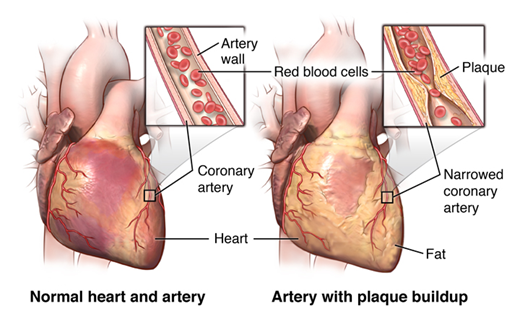
To diagnosis of coronary artery disease, which is the narrowing of the blood vessels that supply oxygen and nutrients to the heart muscle
-
After a heart attack (myocardial infarction, or MI) to look for heart muscle damage
-
To assess blood flow to areas of the heart muscle that have had blood flow restored by bypass surgery, angioplasty, or stent placement
-
To check for scar tissue in the heart from other disease not related to coronary artery disease
There may be other reasons for your healthcare provider to order a resting myocardial perfusion scan.
What are the risks of a resting myocardial perfusion scan?
Except for the needle used to put in the IV, this test does not cause pain.
The injection of the radioactive tracer may cause some slight discomfort. Allergic reactions to the tracer are rare.
You may want to ask your healthcare provider about the amount of radiation used during the procedure and the risks related to your particular situation.
There may be other risks depending on your specific medical condition. Be sure your healthcare provider knows about all of your medical conditions.
Make a list of questions you have about the procedure. Be sure to discuss these questions and any concerns with your healthcare provider before the test. Bring a family member or friend to the appointment to help you remember your questions and concerns.
Certain factors may interfere with or affect the results of this test. These include:
-
Caffeine intake within 48 hours of the procedure
-
Smoking or using any form of tobacco within 48 hours of the procedure
-
Certain heart medicines
How do I prepare for a resting myocardial perfusion scan?
PLEASE NOTE: Certain factors or conditions may interfere with or affect the results of the test. These include, but are not limited to, the following:
-
Caffeine within 24 hours of the procedure
-
Nitrate medications
-
Notify your doctor and when you schedule your exam if you have a pacemaker
PRECAUTIONS: If you are pregnant or think you may be pregnant, please check with your doctor before scheduling the exam. We will discuss other options with you and your doctor.
BREASTFEEDING: If you are breastfeeding, you should notify your health care provider due to the risk of contaminating breast milk with the tracer.
CLOTHING: You may be asked to change into a patient gown. A gown will be provided for you. Lockers are provided to secure your personal belongings. Please remove all piercings and leave all jewelry and valuables at home.
EAT/DRINK: Fasting may be required before the procedure. You will be given specific instructions as to how long you should withhold food and/or liquids when you schedule your appointment. You should refrain from eating or drinking anything that contains caffeine for at least 24 hours prior to the procedure. Some prescription and over-the-counter medications contain caffeine and should be avoided. Some over-the-counter medications that contain caffeien include Anacin, Excedrin and NoDoz.
ALLERGIES: Notify the radiologist or technologist if you are allergic to or sensitive to medications, contrast dyes or iodine. The injection of the radiotracer may cause some slight discomfort. Allergic reactions to the radiotracer are rare, but may occur.
MEDICATIONS: Please bring a list of all medications (prescription and over-the-counter) and herbal supplements that you are taking.
Based on your medical condition, your doctor may request other specific preparation.
What happens during a resting myocardial perfusion scan?
You may have a resting myocardial perfusion scan on an outpatient basis or as part of your stay in a hospital. Procedures may vary depending on your condition and your healthcare providers practice.
Generally, a resting myocardial perfusion scan follows this process:
-
You will be asked to remove any jewelry or other objects that may interfere with the procedure.
-
You will be asked to remove your clothing and will be given a gown to wear.
-
An intravenous (IV) line will be started in your hand or arm.
-
You will be connected to an ECG machine with leads that stick to your skin and a blood pressure cuff will be placed on your arm.
-
You will lie flat on a table in the procedure room.
-
The radioactive tracer will be injected into the IV in your hand or arm.
-
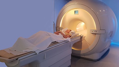
After the tracers has circulated through your body (10 to 60 minutes depending on the type of radioactive tracer being used), the scanner will take pictures of your heart. In this special kind of imaging test, called SPECT (single photon emission computed tomography), the scanner rotates around you as it takes pictures. The table slides into the hole of the scanner, which is a large, donut-shaped machine.
-
You will be lying flat on a table while the images of your heart are made. Your arms will be on a pillow above your head. You will need to lie very still while the images are being taken, as movement can affect the quality of the images.
-
If you have any symptoms, such as dizziness, chest pain, extreme shortness of breath, or severe fatigue, at any point during the procedure, let the healthcare provider know.
-
After the scan is done, the IV line will be removed, and you will be allowed to leave, unless your healthcare provider tells you differently.
What happens after a resting myocardial perfusion scan?
Move slowly when getting up from the scanner table to avoid any dizziness or lightheadedness from lying flat for the length of the procedure.
Drink plenty of fluids and empty your bladder often for 24 to 48 hours after the test. This helps flush the remaining radioactive tracer from your body.
The IV site will be checked for any signs of redness or swelling. If you notice any pain, redness, or swelling at the IV site after you return home, tell your healthcare provider. This may be a sign of infection or other type of reaction.
Your healthcare provider may give your other instructions after the procedure, depending on your particular situation. If the perfusion scan shows you may have a serious or life threatening cardiac disease, your healthcare provider may talk to you about a same-day cardiovascular procedure.