Magnetic Resonance Imaging (MRI)
What You Need to Know
- MRI is a type of diagnostic test that can create detailed images of nearly every structure and organ inside the body.
- MRI uses magnets and radio waves to produce images on a computer. MRI does not use ionizing radiation.
- Images produced by an MRI scan can show organs, bones, muscles and blood vessels.
What is MRI?
Magnetic resonance imaging, or MRI, is a noninvasive medical imaging test that produces detailed images of almost every internal structure in the human body, including the organs, bones, muscles and blood vessels. MRI scanners create images of the body using a large magnet and radio waves. No ionizing radiation is produced during an MRI exam, unlike X-rays. These images give your physician important information in diagnosing your medical condition and planning a course of treatment.
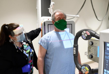
What to Expect During Your MRI Exam at Johns Hopkins Medical Imaging

Learn about the people and technology involved during your MRI exam at Johns Hopkins Medical Imaging. This is meant to be a general overview and we recommend carefully reading your appointment instructions for specific details on how to best prepare for your scheduled exam.
How does an MRI scan work?
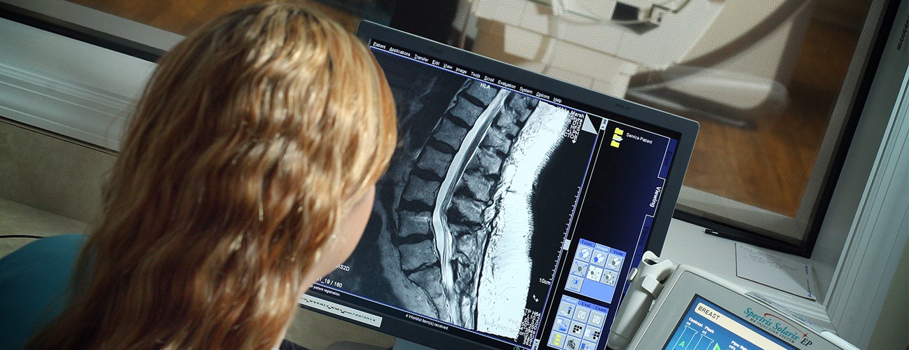
The MRI machine is a large, cylindrical (tube-shaped) machine that creates a strong magnetic field around the patient and sends pulses of radio waves from a scanner. Some MRI machines look like narrow tunnels, while others are more open.
The strong magnetic field created by the MRI scanner causes the atoms in your body to align in the same direction. Radio waves are then sent from the MRI machine and move these atoms out of the original position. As the radio waves are turned off, the atoms return to their original position and send back radio signals. These signals are received by a computer and converted into an image of the part of the body being examined. This image appears on a viewing monitor.
MRI may be used instead of computed tomography (CT) when organs or soft tissue are being studied. MRI is better at telling the difference between types of soft tissues and between normal and abnormal soft tissues.
Because ionizing radiation is not used, there is no risk of exposure to radiation during an MRI procedure.
Newer uses for MRI have contributed to the development of additional magnetic resonance technology. Magnetic resonance angiography (MRA) is a procedure used to evaluate blood flow through arteries. MRA can also be used to detect aneurysms in the brain and vascular malformations — abnormalities of blood vessels in the brain, spinal cord or other parts of the body.
Functional magnetic resonance imaging (fMRI) of the brain is used to determine the specific location in the brain where a certain function, such as speech or memory, occurs. The general areas of the brain in which such functions occur are known, but the exact location may vary from person to person. During fMRI of the brain, you will be asked to perform a specific task, such as reciting the Pledge of Allegiance. By pinpointing the exact location of the functional center in the brain, doctors can plan surgery or other treatments for a brain disorder.
How do I prepare for an MRI procedure?
EAT/DRINK: You may eat, drink and take medications as usual for most MRI exams. There are some specialty MRI exams that require certain restrictions. You will be provided detailed preparations instructions by Johns Hopkins Medical Imaging when you schedule your exam.
CLOTHING: You will be asked to remove all clothing, including underwear, and lock up all personal belongings. Please remove all piercings and leave all jewelry and valuables at home.
WHAT TO EXPECT: Imaging takes place inside of a large, tubelike structure that is open on both ends. You must lie perfectly still for quality images. Due to the MRI machine's loud noise, earplugs are required and will be provided.
ALLERGY: Some MRI exams require IV contrast. If you have had an allergic reaction to MRI contrast, contact your ordering physician to obtain the recommended prescription. You will likely take this by mouth 24, 12 and two hours prior to the examination.
ANTI-ANXIETY MEDICATION: If you require anti-anxiety medication due to claustrophobia, contact your ordering physician for a prescription. You must bring your prescription on the day of your appointment. Please note that you will need someone to drive you home.
STRONG MAGNETIC ENVIRONMENT: Due to the strong magnetic field, you must inform your doctor prior to the appointment if you have any metal in your body. Detailed information will be needed, such as the type and location, to determine your eligibility for MRI. If you have metal in your body that was not disclosed before your appointment, your study may be delayed, rescheduled or canceled upon your arrival until more information can be obtained.
Based on your medical condition, your health care provider may require other preparations.
When you call to make an appointment, it is extremely important that you inform your doctor if any of the following apply to you:
- You have a pacemaker or have had heart valves replaced.
- You have any type of implantable pump, such as an insulin pump.
- You have vessel coils, filters, stents, or clips.
- You are pregnant or think you might be pregnant.
- You have ever had a bullet wound.
- You have ever worked with metal (for example, as a metal grinder or welder).
- You have metallic fragments anywhere in the body.
- You are not able to lie down for 30 to 60 minutes.
How do I prepare for specialized MRI studies?
In some cases, you will be contacted prior to the examination to discuss the details of the procedure and how to prepare.
Specialized MRI exams include:
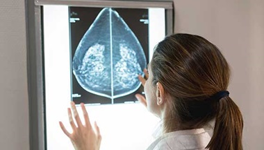
- breast MRI
- breast biopsy by MRI
- dynamic pelvis/defecography by MRI
- enterography by MRI
- functional MRI
- magnetic resonance angiography (MRA)
- prostate imaging by MRI
What happens during an MRI procedure?
MRI scans may be performed on an outpatient basis or as part of a stay in a hospital. Although specific protocols may differ among facilities, an MRI procedure generally follows this process:
- You will be asked to remove all clothing, jewelry, eyeglasses, hearing aids, hairpins, removable dental work or other objects that may interfere with the procedure.
- You will be given a gown to wear.
- If you are to have a procedure done with contrast, an IV line will be started in the hand or arm for injection of the contrast dye. If the contrast is to be taken by mouth, you will be given the contrast to swallow.
- You will lie on a scan table that slides into a large circular opening of the scanning machine. Pillows and straps may be used to prevent movement during the procedure.
- The technologist will be in another room where the scanner controls are located. However, you will be in constant sight of the technologist through a window. Speakers inside the scanner will enable the technologist to communicate with and hear you. You will have a communication ball so that you can let the technologist know if you have any problems during the procedure. The technologist will be watching you at all times and will be in constant communication.
- You will be given earplugs or a headset to help block out the noise from the scanner. Some headsets may play music.
- During the scanning process, a clicking noise will sound as the magnetic field is created and pulses of radio waves are sent from the scanner.
- It will be important for you to remain very still during the examination, as any movement could cause distortion and affect the quality of the scan.
- At intervals, you may be instructed to hold your breath or not breathe for a few seconds, depending on the body part being examined. You will then be told when you can breathe. You should not need to hold your breath for longer than a few seconds.
- If contrast dye is used, you may experience some effects when it is injected into the IV line. These effects include a flushing sensation or a feeling of coldness, a salty or metallic taste in the mouth, a brief headache, itching, or nausea and/or vomiting. These effects usually last for a few moments.
- You should notify the technologist if you have any breathing difficulties, sweating, numbness or heart palpitations.
- Once the scan is complete, the table will slide out of the scanner and you will be helped off the table.
- If an IV line was inserted for contrast administration, the line will be removed.
While the MRI procedure itself causes no pain, having to lie still for the length of the procedure might cause some discomfort or pain, particularly if you had a recent injury or invasive procedure, such as surgery. The technologist will use all possible comfort measures and complete the procedure as quickly as possible.
What happens after an MRI procedure?
You should move slowly when getting up from the scanner table to avoid any dizziness or lightheadedness from lying flat during the procedure.
If any sedatives were taken for the procedure, you may be required to rest until they have worn off. You will also need to avoid driving.
If contrast dye was used during the procedure and you experience any side effects or reactions to the contrast dye, such as itching, swelling, rash or difficulty breathing after your appointment call your doctor right away. If you feel it is a life threatening emergency, call 911.
If you notice any pain, redness and/or swelling at the IV site after you return home, you should notify your doctor, as this could indicate an infection or other type of reaction.
Otherwise, no special type of care is required after an MRI scan. You may resume your usual diet and activities unless your doctor advises you differently.
Your doctor may give you more or alternate instructions after the procedure depending on your particular situation.
If you are a patient at Johns Hopkins Medicine, you should see a final report on your MRI exam in MyChart three or four days after your appointment. (Note: You must sign up for MyChart to have access to your medical records.)
A Child’s MRI without Anesthesia – Johns Hopkins Pediatric Radiology

Is your child about to have an MRI exam? Kids can learn from Fin the fish everything they need to know about coming to Johns Hopkins for an MRI scan. Your child’s MRI exam may or may not use anesthesia. This video explains the process for having a pediatric MRI scan without anesthesia at Johns Hopkins.