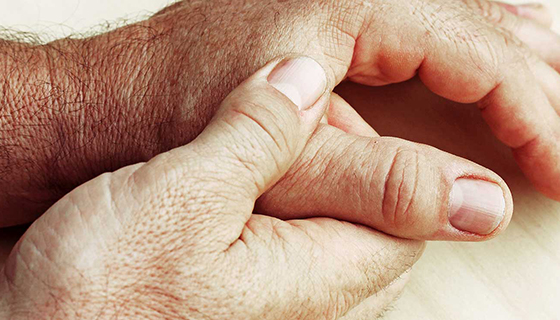
Diagnosing Hand Conditions
How are hand conditions diagnosed?
Some hand conditions need to be diagnosed through surgery. It depends on what is causing the condition. In general, a hand condition may need the following procedures to help in the diagnosis:
-
Complete history and physical exam. Your healthcare provider will need to know your age, hand preference, occupation, and any history of other problems with the hand. For injuries, your healthcare provider may also need to know:
-
Type of injury that occurred
-
When and where the injury occurred
-
Other information about the injury. For example, was it related to work? Was the hand injured with a contaminated piece of machinery or chemical?
-
Position of the thumb or affected fingers during the injury or fall
-
-
Your health history. This includes your tetanus vaccine status and current medicines.
In some cases, a diagnosis can be made simply based on a physical exam. But you may need the following tests to help confirm the diagnosis, or the extent of the problem:
-
Arthrography. A contrast dye is injected into the hand to allow the healthcare provider to better see the joints on X-ray.
-
Bone scan (bone scintigraphy). A slightly radioactive substance (radiotracer, technetium) is injected into a vein, and images are taken to show how the tracer has entered various tissues and structures. The study is often done in phases and may take several hours. Images of the hand are taken at different times after the tracer is injected.
-
CT scan. This test uses X-rays and a computer to make detailed images of the body. It can look at the bones, muscles, fat, and organs. CT scans are more detailed than general X-rays.
-
Electromyogram (EMG). This test measures the electrical activity of a muscle or a group of muscles. An EMG can find abnormal electrical muscle activity caused by diseases and nervous system conditions.
-
MRI. This test uses large magnets, radiofrequencies, and a computer to make detailed images of organs and structures in the body. It lets the healthcare provider see the tendons, ligaments, vessels, and nerves in the hand.
-
Ultrasound (sonography). This imaging test uses high-frequency sound waves and a computer to make images of blood vessels, tissues, and organs. Ultrasounds are used to see internal organs as they function. It can see blood flow through various vessels. In the hand, ultrasound is useful for finding fluid collections, such as cysts.
-
Video fluoroscopy. This test lets your healthcare provider see how your hand moves by recording the movement with an X-ray video for repeated viewing. The image is displayed on a screen.
-
X-ray. This test uses electromagnetic radiation to make images of internal tissues, bones, and organs.