Craniosynostosis
What You Need to Know
- Craniosynostosis is common and occurs in one out of 2,200 live births.
- The condition affects males slightly more often than females.
- Craniosynostosis is most often sporadic (occurs by chance) but can be inherited in some families.
What is craniosynostosis?
In fetuses and newborns, the skull consists of several plates of bone that are separated by flexible, fibrous joints called sutures. As infants grow and develop, the sutures close, forming a solid piece of bone.
Craniosynostosis is a condition in which the sutures close too early, causing problems with normal brain and skull growth. Premature closure of the sutures may also cause pressure inside the head to increase and the skull or facial bones to change from a normal, symmetrical appearance.
What causes craniosynostosis?
Craniosynostosis is a feature of many different genetic syndromes that have a variety of inheritance patterns and chances for recurrence, depending on the specific syndrome present.
It is important for the child with craniosynostosis and his/her family members to be examined carefully for signs of an inherited genetic disorder, such as limb defects, ear abnormalities or heart defects.
Craniosynostosis Symptoms
In infants with this condition, the most common signs are changes in the shape of the head and face. One side of your child’s face may look markedly different from the other side. Other, much less common signs may include:
- A full or bulging fontanelle (soft spot located on the top of the head)
- Sleepiness (or less alert than usual)
- Very noticeable scalp veins
- Increased irritability
- High-pitched cry
- Poor feeding
- Projectile vomiting
- Increasing head circumference
- Developmental delays
The symptoms of craniosynostosis may resemble other conditions or medical problems, so always work with your child’s physician to clarify a diagnosis.
Different Types of Craniosynostosis
Brachycephaly
Anterior brachycephaly involves fusion of either the right or left side of the coronal suture that runs across the top of the baby’s head from ear to ear.
This is called coronal synostosis, and it causes the normal forehead and brow to stop growing. The result is a flattening of the forehead and the brow on the affected side, with the forehead tending to be excessively prominent on the opposite side. The eye on the affected side may also have a different shape, and there may be flattening of the back of the head (occipital). When the suture fusion is all the way across the back of the child’s skull, the result is posterior plagiocephaly.
Trigonocephaly
Trigonocephaly is a fusion of the metopic (forehead) suture. This suture runs from the top of the head down the middle of the forehead, toward the nose.
Early closure of this suture may result in a prominent ridge running down the forehead. Sometimes, the forehead looks quite pointed, like a triangle, with closely placed eyes (hypotelorism).
Scaphocephaly
Scaphocephaly is an early closure or fusion of the sagittal suture. This suture runs front to back, down the middle of the top of the head. This fusion causes a long, narrow skull. The skull is long from front to back and narrow from ear to ear.

Craniosynostosis Diagnosis
Craniosynostosis may be congenital (present at birth) or observed later, often during a physical examination in the first year of life.
The diagnosis involves thorough physical examination and diagnostic testing. Your child’s doctor will start with a complete prenatal and birth history, asking about any family history of craniosynostosis or other head or face abnormalities.
The doctor may also ask about developmental milestones, since craniosynostosis can be associated with other neuromuscular disorders. Developmental delays may require further medical follow-up for underlying problems.
During the examination, the doctor will measure the circumference of your child’s head to identify normal and abnormal ranges. Craniosynostosis can be diagnosed by physical exam. If needed, your neurosurgeon may recommend imaging tests.
Craniosynostosis: Fitz’s Story
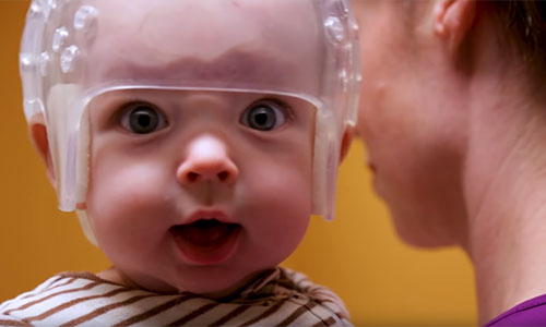
When Fitz was born, it was obvious that his skull was misshapen. By 5 weeks old, Fitz had been diagnosed with craniosynostosis. His skull had fused early and was constricting his brain growth.
Craniosynostosis Treatment
The key to treating craniosynostosis is early detection and treatment. Specific therapy for craniosynostosis will be determined by your child’s physician based on:
- Your child’s age, overall health and medical history
- Extent of the craniosynostosis
- Type of craniosynostosis (which sutures are involved)
- Your child’s tolerance for specific medications, procedures or therapies
- Expectations for the course of the craniosynostosis
- Your opinion or preference
Surgery is typically the recommended treatment, since it can reduce pressure in the head and correct the deformities of the face and skull bones.
Early diagnosis and consultation with a specialist are important. In general, the best time to operate is before the child is 1 year old, since the bones are still very soft and easy to work with. If your child’s condition is severe, the doctor may recommend surgery as early as 1 month of age.
Before surgery, your child’s physician will explain the operation and may review before-and-after photographs of children who have had a similar type of surgery.
Calvarial Vault Remodeling
In this procedure, the surgeon makes an incision in the infant’s scalp and corrects the shape of the head by moving the area of the skull that is abnormally or prematurely fused, and then reshapes the skull so it can take more of a round contour. Surgery can last up to six hours. Your baby will likely spend one night in the intensive care unit, plus an additional few days in the hospital for monitoring.
Even if your child’s deformity is seen early on, this surgery is best suited for babies 5-6 months of age or older to ensure the bone is thick enough to perform the needed reshaping. This surgery may commonly involve a blood transfusion.
After surgery, there may be temporary facial swelling. Unlike other surgical options, there are no additional steps post-surgery unless a recurrence of craniosynostosis is found. You can expect to follow up with your surgery team one month post-surgery to check on the surgery incision site, and again at six and 12 months after the procedure to ensure healing is progressing.
Endoscopic Craniosynostosis Surgery
Some hospitals may offer the option of this minimally invasive surgery, which may be performed when the baby is 2–3 months old, depending on the type and degree of craniosynostosis.
The procedure involves the use of an endoscope, a small tube that the surgeon can look through and see immediately inside and outside the skull through very small incisions in the scalp. The surgeon opens the prematurely fused suture to enable the baby’s brain to grow normally.
The surgery itself takes approximately one hour and involves less blood loss compared with cranial vault remodeling, so there is less chance of requiring a blood transfusion. Your baby will stay in the hospital overnight for monitoring before being released to go home.
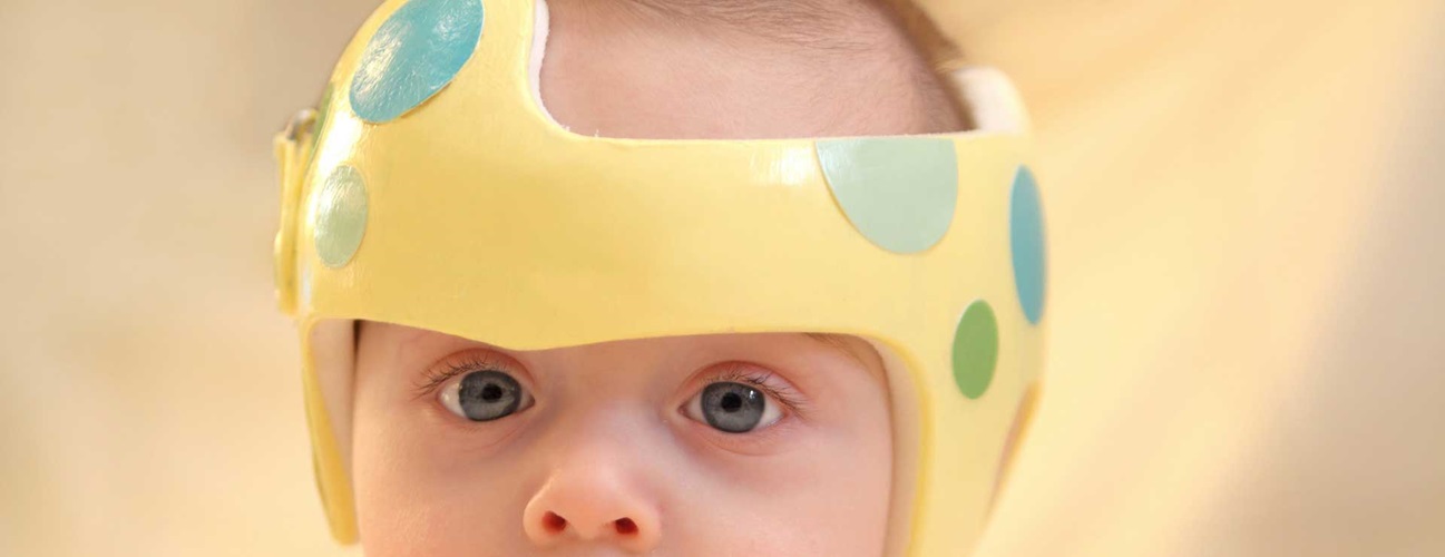
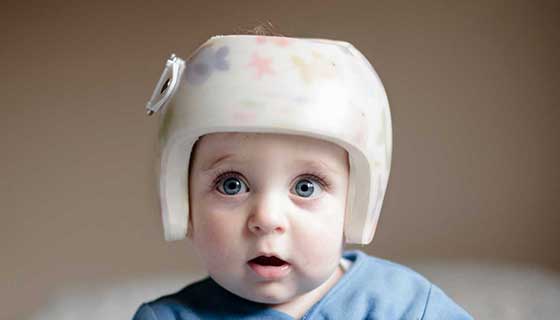
This type of surgery is followed by the use of a molding helmet to reshape the skull. Additional appointments with the helmet provider (orthotist) will be necessary for fitting the helmet to your child. You can expect to follow up with your surgery team every three months for the first year post-surgery to check progress of the skull reshaping.
After Craniosynostosis Surgery
Following craniosynostosis surgery, your child will likely have a turbanlike dressing around his or her head, and may experience swelling in the face and eyelids. Your child will spend the period after surgery in an intensive care unit for close monitoring.
The care team will watch closely for any problems after surgery, such as:
- Fever (greater than 101 degrees Fahrenheit)
- Vomiting
- Irritability
- Redness and swelling along the incision areas
- Decreased alertness
These complications require prompt evaluation by your child’s surgeon.
Follow-Up Care
The recovery process is different for each child. Your child’s health care team will work with your family, giving you instructions on how to care for your child at home and outlining specific problems that require immediate medical attention.
Craniosynostosis can affect a child’s brain and development. The degree of the problems depends on the severity of the craniosynostosis, the number of sutures that are fused, and the presence of brain or other organ system problems that could affect the child.
The physician may recommend genetic counseling to evaluate the child’s parents for any disorders that may run in families.
A child with craniosynostosis requires frequent medical evaluations to ensure that the skull, facial bones, jaw alignment and brain are developing normally. The medical team will provide education and guidance to help you make the most of your child’s health and well-being.
The Johns Hopkins Cleft and Craniofacial Center
