Ulcerative Colitis
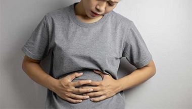
Ulcerative colitis is an inflammatory bowel disease in which the inner lining of the large intestine and rectum become inflamed. Ulcerative colitis is characterized by diarrhea, abdominal pain and blood in the stool. The disease may vary in how much of the colon is affected and in severity as well.
The difference between ulcerative colitis and Crohn’s disease is that the inflammation only occurs on the inner lining of the bowel in ulcerative colitis, while the inflammation can affect all layers of the bowel from Crohn’s disease.

Ulcerative Colitis Symptoms
Symptoms may include:
-
Bloody diarrhea, often the main symptom of ulcerative colitis
-
Frequent bowel movements
-
Abdominal or rectal pain
-
Fever
-
Weight loss
-
Joint pain
-
Skin rashes
-
Occasionally, constipation and rectal spasm
Ulcerative Colitis Diagnosis
A thorough physical examination and careful monitoring help obtain an accurate diagnosis. A doctor will monitor before and during an ulcerative colitis attack with blood work and a colonoscopy. The colonoscopy gives the care team valuable information by noting the length and extent of the attack.
Another good indicator of the severity of the disease is how often and how loose and bloody the diarrhea is. A doctor will especially look to see if the frequency of bowel movements increases during an attack.
The doctor will also order blood tests to help confirm a diagnosis. Other diagnostic procedures include:
Imaging Scans
Imaging scans allow your doctor to get detailed images of the affected area. Imaging studies include:
-
Computed tomography (CT) scan: A CT scan is a powerful X-ray that is very useful in diagnosing ulcerative colitis. It provides accurate images of the bowel wall.
-
Barium enema: A barium preparation is inserted through a rectal tube, coating the entire colon. X-rays are taken of the colon.
Flexible Sigmoidoscopy
If you have lower abdominal symptoms, you may need a flexible sigmoidoscopy. A sigmoidoscopy is a type of endoscopic procedure that allows your doctor to examine part of your colon from the rectum. The test takes 10 to 20 minutes.
A flexible sigmoidoscopy examines the rectum and lower colon. During the procedure:
-
Your colon must be clear of stool so your doctor has good visibility. Preparations may include a liquid diet, enema and laxatives.
-
Your doctor inserts the sigmoidoscope, a thin, flexible tube, through the rectum and into the anus and large intestine to view the area.
-
The procedure may cause some cramping or discomfort.
The Importance of Good Bowel Preparation During Colonoscopy
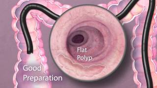
In this video, learn why the bowel preparation for a colonoscopy is so important to the results of the procedure.
Colonoscopy
A colonoscopy is similar to a flexible sigmoidoscopy but takes longer (30 to 60 minutes) and allows your doctor to examine the entire large intestine.
During a colonoscopy:
-
Your colon must be clear of stool so your doctor has good visibility. Preparations may include a liquid diet, enema and laxatives.
-
You are sedated before the procedure.
-
Your doctor inserts the colonoscope through the rectum and into the anus and large intestine.
-
A biopsy forceps may be inserted through the scope in order to remove a small sample of tissue for further analysis.
-
The procedure may cause some cramping or discomfort.