Cerebral Aneurysm
What You Need to Know
- A brain aneurysm (also called a cerebral aneurysm or an intracranial aneurysm) is a ballooning arising from a weakened area in the wall of a blood vessel in the brain.
- If the brain aneurysm expands and the blood vessel wall becomes too thin, the aneurysm will rupture and bleed into the space around the brain. This event is called a subarachnoid hemorrhage (SAH) and may cause a hemorrhagic (bleeding) stroke.
- Rupturing brain aneurysm and SAH are life-threatening events. If you suspect you are having a rupturing brain aneurysm, call 911.
What are the symptoms of brain aneurysm?
The presence of a brain aneurysm may not be known until it ruptures. Most brain aneurysms have no symptoms and are small in size (less than 10 millimeters, or less than four-tenths of an inch, in diameter). Smaller aneurysms may have a lower risk of rupture.
However, occasionally there may be symptoms that happen before a rupture due to a small amount of blood that may leak. This is called "sentinel hemorrhage" into the brain. Some aneurysms are symptomatic because they press on adjacent structures, such as nerves to the eye. They can cause visual loss or diminished eye movements, even if the aneurysm has not ruptured.
The symptoms of an unruptured brain aneurysm include the following:
-
Headaches (rare, if unruptured)
-
Eye pain
-
Vision changes
-
Diminished eye movement
The first evidence of a brain aneurysm is most often a subarachnoid hemorrhage (SAH), due to rupture of the aneurysm. This may cause symptoms such as:
-
Rapid onset of "worst headache of my life"
-
Stiff neck
-
Nausea and vomiting
-
Changes in mental status, such as drowsiness
-
Pain in specific areas, such as the eyes
-
Dilated pupils
-
Loss of consciousness
-
High blood pressure
-
Loss of balance or coordination
-
Sensitivity to light
-
Back or leg pain
-
Problems with certain functions of the eyes, nose, tongue, and/or ears that are controlled by one or more of the 12 cranial nerves
-
Coma and death
The symptoms of a brain aneurysm may resemble other problems or medical conditions. Always talk with your healthcare provider for a diagnosis.
What are the risk factors for brain aneurysm?
Inherited risk factors associated with aneurysm formation may include the following:
-
Alpha-glucosidase deficiency. A complete or partial deficiency of the enzyme needed to break down glycogen and to convert it into glucose.
-
Alpha 1-antitrypsin deficiency. A hereditary disease that may lead to hepatitis and cirrhosis of the liver or emphysema of the lungs.
-
Arteriovenous malformation (AVM). An abnormal connection between an artery and a vein.
-
Coarctation of the aorta. A narrowing of the aorta. This is the main artery coming from the heart.
-
Ehlers-Danlos syndrome. A connective tissue disorder (less common).
-
Family history of aneurysms
-
Female gender
-
Fibromuscular dysplasia. An arterial disease, cause unknown, that most often affects the medium and large arteries of young to middle-aged women.
-
Hereditary hemorrhagic telangiectasia. A genetic disorder of the blood vessels in which there is a tendency to form blood vessels that lack capillaries between an artery and vein.
-
Klinefelter syndrome. A genetic condition in men in which an extra X sex chromosome is present.
-
Noonan's syndrome. A genetic disorder that causes abnormal development of many parts and systems of the body.
-
Polycystic kidney disease (PCKD). A genetic disorder characterized by the growth of numerous cysts filled with fluid in the kidneys. PCKD is the most common medical disease associated with saccular aneurysms.
-
Tuberous sclerosis. A type of neurocutaneous syndrome that can cause tumors to grow inside the brain, spinal cord, organs, skin, and skeletal bones.
Acquired risk factors associated with aneurysm formation may include the following:
-
Advancing age
-
Alcohol consumption (especially binge drinking)
-
Atherosclerosis. A buildup of plaque (made up of deposits of fatty substances, cholesterol, cellular waste products, calcium, and fibrin) in the inner lining of an artery
-
Cigarette smoking
-
Use of illicit drugs, such as cocaine or amphetamine
-
High blood pressure
-
Head injury
-
Infection
Although these risk factors increase a person's risk, they do not necessarily cause the disease. Some people with one or more risk factors never develop the disease, while others develop disease and have no known risk factors. Knowing your risk factors to any disease can help to guide you into the appropriate actions. These include changing behaviors and being monitored for the disease.
Brain Aneurysm Diagnosis
A brain aneurysm is often discovered after it has ruptured or by chance during diagnostic exam, such as computed tomography (CT scan), magnetic resonance imaging (MRI), or angiography that are being done for other reasons.
In addition to a complete medical history and physical exam, diagnostic procedures for a brain aneurysm may include:
-
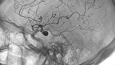
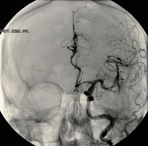
Cerebral angiography. This provides an image of the blood vessels in the brain to detect a problem with vessels and blood flow. The procedure involves inserting a catheter (a small, thin tube) into an artery in the leg and passing it up to the blood vessels in the brain. Contrast dye is injected through the catheter and X-ray images are taken of the blood vessels.
-
Computed tomography scan (CT or CAT scan). This is an imaging test that uses X-rays and a computer to make detailed images of the body. A CT scan shows details of the bones, muscles, fat, and organs. CT scans are more detailed than general X-rays and may be used to detect abnormalities and help identify the location of the aneurysm and if it has burst or is leaking. A CT angiogram (CTA) can also be obtained on a CT scan to look at the vessels.
-
Magnetic resonance imaging (MRI). A diagnostic procedure that uses a combination of large magnets, radiofrequencies, and a computer to produce detailed images of organs and structures within the body. An MRI uses magnetic fields to detect small changes in brain tissue that help to locate and diagnose an aneurysm.
-
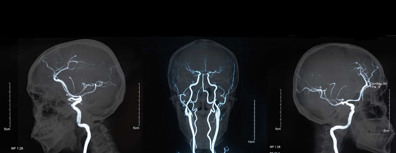
Magnetic resonance angiography (MRA). A noninvasive diagnostic procedure that uses a combination of magnetic resonance technology (MRI) and intravenous (IV) contrast dye to visualize blood vessels. Contrast dye causes blood vessels to appear opaque on the MRI image, allowing the doctor to visualize the blood vessels being evaluated.
Brain Aneurysm Treatment
Brain aneurysms are treated using one or more of the following methods, depending on the location and size of the aneurysm and whether or not it has ruptured, as well as the individual patient’s needs:
Brain Aneurysms: FAQs with Raphael Tamargo, M.D.
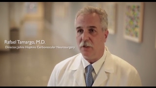
Neurosurgeon Rafael Tamargo, M.D., Director of Johns Hopkins' Aneurysm Center, explains what a brain aneurysm is, what steps to take if you have this diagnosis, available surgical options and anticipated outcomes from each surgical option.
Dr. Olachi Mezu suffered — and survived — a ruptured brain aneurysm

Dr. Olachi Mezu suffered a ruptured brain aneurysm while traveling from New York to her home in Maryland. Watch her story as she talks about the care and treatment she received from Johns Hopkins and her neurosurgeon Dr. Judy Huang.