Aneurysm
What is an aneurysm?
An aneurysm is a bulging, weakened area in the wall of a blood vessel resulting in an abnormal widening or ballooning greater than 50% of the vessel's normal diameter (width). An aneurysm may occur in any blood vessel, but is most often seen in an artery rather than a vein.
An aneurysm may be located in many areas of the body, such as blood vessels of the brain (cerebral aneurysm), the aorta (the largest artery in the body), the neck, the intestines, the kidney, the spleen, and the vessels in the legs (iliac, femoral, and popliteal aneurysms). The most common location of an aneurysm is the aorta, which carries oxygenated blood from the heart to the body. The thoracic aorta is the short segment of the aorta in the chest cavity. The abdominal aorta is the section of the aorta that runs through the abdomen. An aneurysm can be characterized by its location, shape, and cause.
The shape of an aneurysm is described as being fusiform or saccular, which helps to identify a true aneurysm. The more common fusiform-shaped aneurysm bulges or balloons out on all sides of the blood vessel. A saccular-shaped aneurysm bulges or balloons out only on one side.
")
A pseudoaneurysm, or false aneurysm, is not an enlargement of any of the layers of the blood vessel wall. A false aneurysm may be the result of a prior surgery or trauma. Sometimes, a tear can occur on the inside layer of the vessel. As a result, blood fills in between the layers of the blood vessel wall creating a pseudoaneurysm.
A dissecting aneurysm is an aneurysm that occurs with a tear in the artery wall that separates the 3 layers of the wall, rather than ballooning out the entire wall.
Because an aneurysm may continue to increase in size, along with progressive weakening of the artery wall, surgical intervention may be needed. Preventing rupture of an aneurysm is 1 of the goals of therapy. The larger an aneurysm becomes, the greater the risk for rupture (bursting). With rupture, life-threatening hemorrhage (uncontrolled bleeding), and possibly death, may result.
What causes an aneurysm to form?
An aneurysm may be caused by multiple factors that result in the breaking down of the well-organized structural components (proteins) of the aortic wall that provide support and stabilize the wall. The exact cause isn't fully known. Atherosclerosis (hardening of the arteries with a sticky substance called plaque) is thought to play an important role in aneurysmal disease. Risk factors associated with atherosclerosis include, but are not limited to:
-
Older age
-
Male
-
Family history
-
Genetic factors
-
Hyperlipidemia (elevated fats and cholesterol in the blood)
-
Smoking
Other specific causes of aneurysms are related to the location of the aneurysm. Examples of aneurysms in the body and their additional causes may include, but are not limited to:
Abdominal Aortic Aneurysm (AAA)
- Atherosclerosis (especially in the segment of the abdominal aorta below the kidneys, called an infrarenal aortic aneurysm)
- Genetic disorders
- Giant cell arteritis (a disease that causes inflammation of the temporal arteries and other arteries in the head and neck, causing the arteries to narrow, reducing blood flow in the affected areas; may cause persistent headaches and vision loss)
- Infection
Cerebral Aneurysm
- Congenital (present at birth)
- High blood pressure
- Atherosclerosis
- Head trauma
Common Iliac Artery Aneurysm
- Atherosclerosis
- Pregnancy
- Infection
- Trauma after lumbar or hip surgery
Femoral and Popliteal Artery Aneurysm
- Atherosclerosis
- Trauma
- Congenital disorders
What are the symptoms of an aneurysm?
Aneurysms may be asymptomatic (no symptoms) or symptomatic (with symptoms). Symptoms associated with aneurysms depend on the location of the aneurysm in the body.
Symptoms that may occur with different types of aneurysms may include, but are not limited to:
- Abdominal Aortic Aneurysm (AAA): Constant pain in abdomen, chest, lower back, or groin area
- Cerebral Aneurysm: Sudden severe headache, nausea, vomiting, visual disturbance, loss of consciousness
- Common Iliac Aneurysm: Lower abdominal, back, and/or groin pain
- Femoral and Popliteal Artery Aneurysm: Easily palpated (felt) pulsation of the artery located in the groin area (femoral artery) or on the back of the knee (popliteal artery), pain in the leg, sores on the feet or lower legs
The symptoms of an aneurysm may resemble other medical conditions or problems. Always consult your doctor for more information.
How are aneurysms diagnosed?
Selection of a type of diagnostic examination is related to the location of the aneurysm. In addition to a complete medical history and physical examination, diagnostic procedures for an aneurysm may include any, or a combination, of the following:
-
Computed tomography (CT or CAT) scan. This diagnostic imaging procedure uses a combination of X-rays and computer technology to produce horizontal, or axial, images (often called slices) of the body. A CT scan shows detailed images of any part of the body, including the bones, muscles, fat, and organs. CT scans are more detailed than standard X-rays.
-
Magnetic resonance imaging (MRI). An MRI uses a combination of large magnets, radio frequencies, and a computer to produce detailed images of organs and structures within the body.
-
Echocardiogram (echo). This procedure evaluates the structure and function of the heart by using sound waves recorded on an electronic sensor that produce a moving picture of the heart and heart valves.
-
Arteriogram (angiogram). This is an X-ray image of the blood vessels used to evaluate various conditions, such as aneurysm, stenosis (narrowing of the blood vessel), or blockages. A dye (contrast) will be injected through a thin flexible tube placed in an artery. This dye will make the blood vessels visible on the X-ray.
-
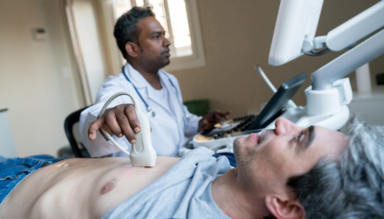
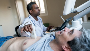
Ultrasound. An ultrasound uses high-frequency sound waves and a computer to create images of blood vessels, tissues, and organs. An ultrasound is used to view internal organs as they function, and to assess blood flow through various vessels.
What is the treatment for aneurysms?
Specific treatment will be determined by your doctor based on:
-
Your age, overall health, and medical history
-
Extent of the disease (location, size, and growth rate of the aneurysm)
-
Your signs and symptoms
-
Your tolerance of specific medications, procedures, or therapies
-
Expectations for the course of the disease
-
Your opinion or preference
Treatment options for an aneurysm may include one or more of the following:
-
Routine ultrasound procedures. These procedures will monitor the size and rate of growth of the aneurysm every 6 months to 12 months as part of a "watchful waiting" approach for smaller aneurysms.
-
Controlling or modifying risk factors. Steps such as quitting smoking, controlling blood sugar if diabetic, losing weight if overweight or obese, and controlling dietary fat intake may help to control the progression of the aneurysm.
-
Medication. Medication can control factors such as hyperlipidemia (elevated levels of fats and cholesterol in the blood) and/or high blood pressure.
-
Surgery:
-
Aneurysm open repair. An incision is made to directly visualize and repair the aneurysm. A cylinder-like tube called a graft may be used to repair the aneurysm. Grafts are made of various materials, such as Dacron (textile polyester synthetic graft) or polytetrafluoroethylene (PTFE, nontextile synthetic graft). This graft is sewn to the involved blood vessel, connecting 1 end of the artery at the site of the aneurysm to the other end. The open repair is considered the surgical standard for an abdominal aortic aneurysm repair
-
Endovascular aneurysm repair (EVAR). EVAR is a procedure that requires only small incisions in the groin along with the use of X-ray guidance and specially-designed instruments to repair the aneurysm. With the use of special endovascular instruments and X-ray images for guidance, a stent-graft is inserted via the femoral artery and advanced up into the aorta to the site of the aneurysm. A stent-graft is a long cylinder-like tube made of thin metal mesh framework (stent), while the graft is made of various materials, such as Dacron or PTFE. The graft material may cover the stent. The stent helps to hold the graft open and in place.
-
#TomorrowsDiscoveries: Therapy for Aortic Aneurysms—Dr. Hal Dietz

#TomorrowsDiscoveries: Johns Hopkins researchers have identified the genes responsible for aortic ballooning and the sequence of events leading to aortic aneurysms. Dr. Hal Dietz currently conducts clinical trials of therapies for people with inherited aortic aneurysms to improve health and quality of life for these patients.