Pancreatic Cancer Diagnosis
Reviewed By:

Tumors of the pancreas are extremely difficult to diagnose because the organ sits deep in the abdomen and is hidden behind other organs. Several diagnostic techniques, including imaging tests and blood tests, may be performed to determine if there is a tumor in the pancreas.
Although various imaging techniques may reveal a mass in the pancreas, the most accurate way to diagnose pancreatic cancer is by studying a biopsied tissue sample under the microscope. Understanding the stage (severity) of the tumor is key to choosing the best treatment.
Computerized Tomography (CT) Scan
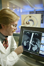
This is an imaging test that combines special X-ray equipment with sophisticated computers to produce multiple images of the inside of the abdomen. It is very useful in detecting the spread of pancreatic cancer to the liver or nearby lymph nodes. CT scans are often performed to monitor patients after treatment to determine whether the cancer has recurred, changed in size or metastasized (spread elsewhere in the body).
Positron Emission Tomography (PET) Scan
For this nuclear medicine test, a small amount of radioactive sugar is injected through a vein before the body is scanned. The radioactive sugar collects mainly in cancer cells, which then show up on the images. This test is not as specific as CT scanning and is not used alone to diagnose pancreatic cancer. A PET scan is often done in combination with a CT scan.
Magnetic Resonance Imaging (MRI)
MRI uses radiofrequency waves and a strong magnetic field rather than X-rays to provide remarkably clear and detailed pictures of internal organs and tissues. This technique has proven very valuable for the diagnosis of a broad range of conditions, including cancer.
Endoscopic Ultrasound (EUS) and Fine Needle Biopsy
During this procedure, the doctor passes a thin, lighted tube called an endoscope through the patient's mouth, down through the stomach and into the first part of the small intestine. At the tip of the endoscope is a device that uses ultrasound waves that produce patterns of echoes as they bounce off internal organs. These ultrasonic patterns can help identify small cancers that cannot be detected by a CT scan. Using X-ray or ultrasound techniques to help guide the needle, the doctor inserts a very thin needle into the pancreas to remove cells to be studied under the microscope.
Endoscopic Retrograde Cholangiopancreatography (ERCP)
Using this technique, the doctor passes the endoscope through the patient's mouth, down through the stomach and into the first part of the small intestine. A smaller catheter tube is then inserted through the endoscope into the bile ducts and pancreatic ducts. Dye is injected through the catheter into the ducts, allowing X-rays to capture pictures that show whether the ducts are narrowed or blocked by a tumor.
Percutaneous Transhepatic Cholangiography (PTC)
This technique is used to take pictures of the bile ducts that drain the liver. Dye is injected through a thin needle inserted through the skin and into the liver, allowing X-ray images to be taken. Unless there is a blockage, the dye should move freely through the bile ducts. From the pictures, the doctor can tell whether there is a blockage from a tumor or other condition. Due to the invasive nature of this procedure, it is only done if ERCP cannot be performed.
CA 19-9 Blood Test
When used with other tests, this tumor marker blood test can assist with the initial diagnosis of pancreatic cancer. In evaluating treatments, CA 19-9 levels may indicate treatment efficacy and the progression of the disease. Since other types of cancer and noncancerous conditions may also lead to elevated CA 19-9 levels, tumor marker test results should be analyzed carefully along with other diagnostic methods.


