Images of our Work:
![]()
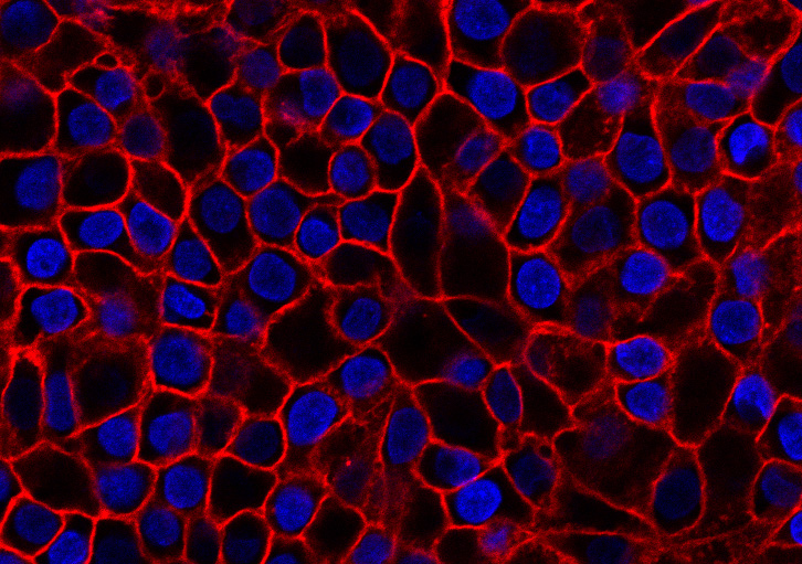
Delivery of photoaffinity & click tagged ganglioside GM1 to human epithelial cells in culture.
Red: Alexa 555 click tag; Blue: nuclei
![]()
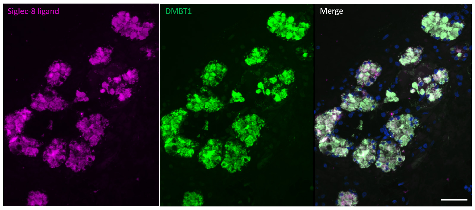
Siglec-8 ligand on human airways is a glycoform of DMBT1. Serial cross section of human airway
submucosal gland was double-stained with Siglec-8-Fc (violet) and anti-DMBT1 antibody (green). The
overlay (right) shows the molecules are congruent. Scale bar, 50 µm. The combined image includes
a marker for cell nuclei (blue)
![]()
Yu, et al., Glycobiology 27, 657-668 (2017)
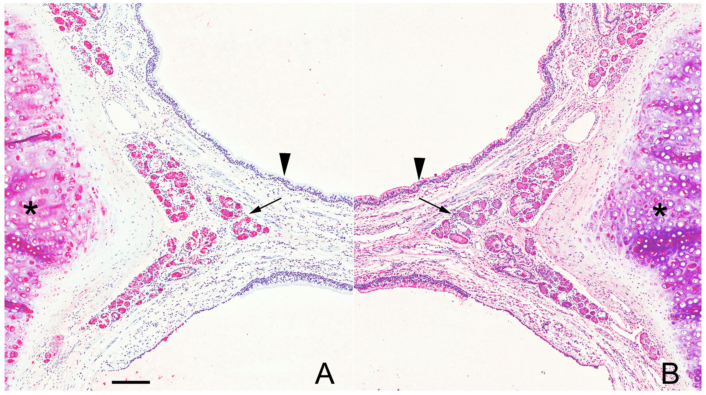
Siglec-8 (A) and Siglec-9 (B) ligands on human bronchus. Serial cross sections of human airway
at the junction of the primary and secondary bronchus were stained with Siglec-8-Fc (A)
or Siglec-9-Fc (B). Images are displayed in mirror image to emphasize differential siglec ligand
distributions. Arrowheads: airway epithelium; arrows: submucosal glands; asterisks: cartilage.
Scale bar, 200 µm.
![]()
Jia, et al., J Allergy Clin Immunol 135, 799-810 (2015)
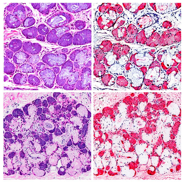
Human tracheal histology (H&E, left panels) stained for Siglec-8 ligands (right top) and Siglec-9 ligands (right bottom).
Siglec-8 ligands are found in serosal cells and ducts of the submucosal glands, whereas Siglec-9 ligands are in
serosal cells, ducts, connective tissue, and (light staining) mucosal cells.
Dorsal root ganglion neurites (labeled with anti-tubulin
antibody) were analyzed for length and integrity. Healthy neurites Left: Dorsal root ganglion neurons labelled with anti-tubulin
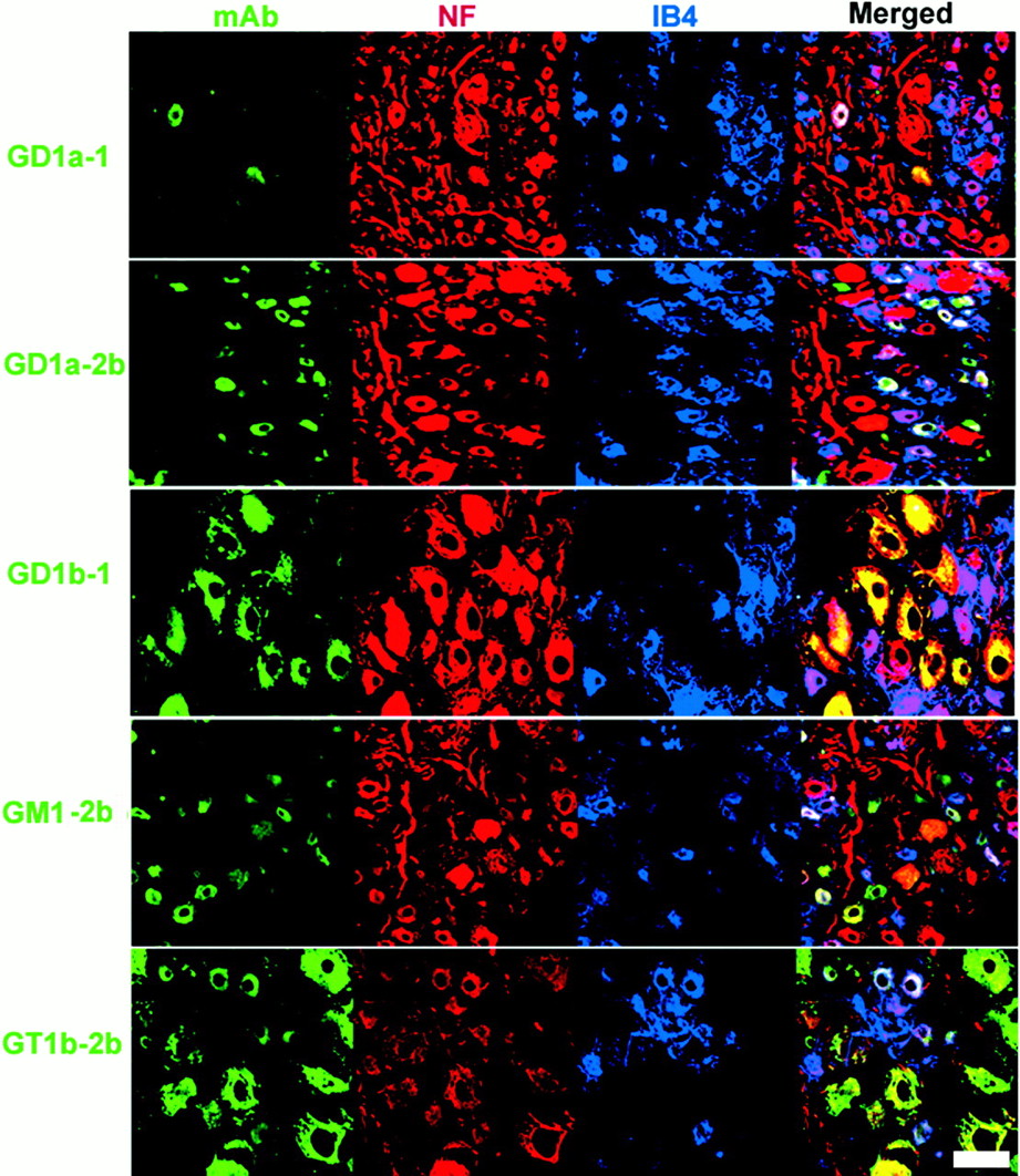
antibody. Top, control; Bottom, inhibited by myelin-associated Dorsal root ganglia triple-labelled with anti-ganglioside mAbs (green), neurofilament (red) and IB-4 (blue). 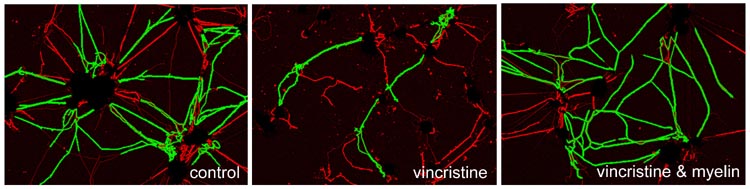 Mehta, et al., ACS Chemical Neuroscience 1,
215-222 (2010)
Mehta, et al., ACS Chemical Neuroscience 1,
215-222 (2010)
are green and segmented (degenerating) neurites are red. The chemotherapeutic
agent vincristine causes degeneration
(center panel). Myelin protects neurites from vincristine toxicity (right
panel).![]()

Mehta, et al., J Biol Chem 282, 27875-27886 (2007)
glycoprotein (MAG).Right: MAG inhibits axon outgrowth via two independent pathways.![]()
Gong, et al., Brain 125, 2491-2506 (2002)
Co-localization of three labels is also shown (Merged). The mAbs included in this figure are shown on the left. Bar = 20 µm.![]()
Lunn, et al., J. Neurochem. 75, 404-412 (2000)
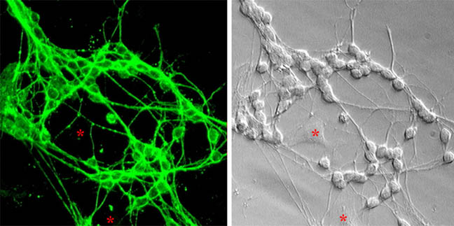
Gangliosides on neurons in cell culture. Rat cerebellar
granule neurons immunostained with anti-GD1a antibody (left).
Corresponding Hoffman modulation contrast image (right). Asterisk denotes
non-neuronal cells, which fail to stain.
![]()
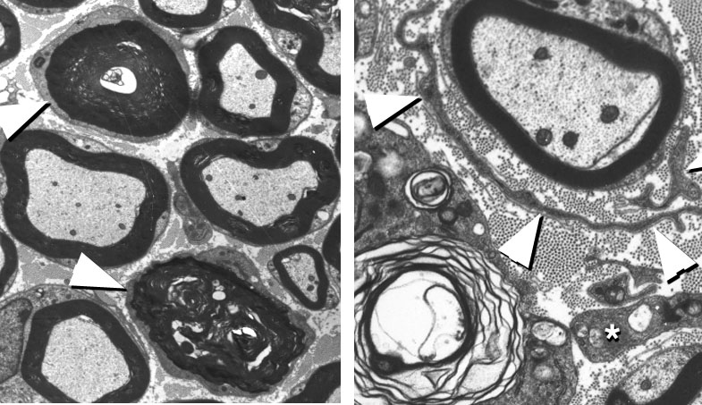
Axon degeneration (left) and demyelination (right) in "complex ganglioside" knockout mice
![]()
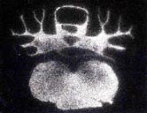
Binding of radiolabeled ganglioside ligand to receptors in the rat brain. Left: Rat brain section stained Confocal immunolocalization of the hepatic lectin on the surface of rat hepatocytes
Return to HOME PAGE or go to: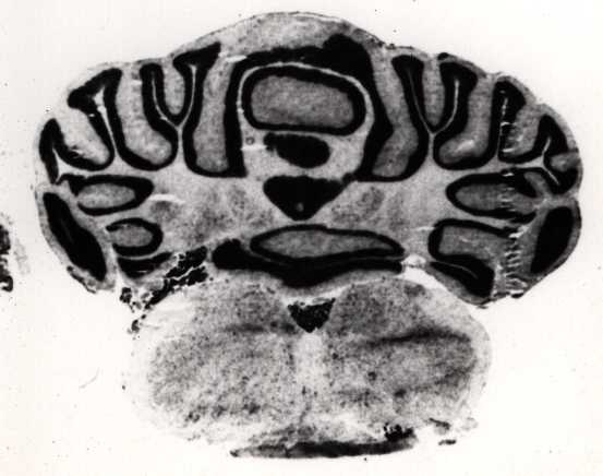
Tiemeyer, et al., J. Biol. Chem. 265, 11990-11999 (1990).
with cresyl violet. Right: Adjacent section treated with 125I-GT1b-BSA, washed, and subjected to autoradiography.
Note prominent white matter but not gray matter distribution of ganglioside receptors. ![]()
Weisz and Schnaar, J. Cell Biol. 115, 485-493 (1991)
adhering to galactose-derivatized surfaces![]()