-
Dara L. Kraitchman, V.M.D., Ph.D., M.S.
- Cardiovascular Interventional Section Head
- Center for Image-Guided Animal Therapy Director
- Professor of Radiology and Radiological Science
- Professor of Molecular and Comparative Pathobiology
MR Sections
Our Research Areas
Cardiac and Interventional MR
The Cardiac and Interventional MR section has projects to develop miniaturized MR detectors on guidewires for endoscopic MRI at ≤100μm resolution and precision-guided high-frequency intravascular ultrasound ablation. High-speed intravascular MRI methods for imaging vessel disease at ~10 fr/sec are being developed, along with precision MRI-guided interventional procedures that could treat obesity and cancer. Ongoing MR spectroscopy (MRS) studies of cardiac energetics in heart failure patients are underway in collaboration with investigators in the Division of Cardiology.
Our Team

The Cellular Imaging Section
The Cellular Imaging Section is located in the Institute for Cell Engineering. We engineer cells so they can be detected with non-invasive imaging techniques such as magnetic resonance imaging and magnetic particle imaging. Our primary mission is to develop new magnetic tracers and cell labeling procedures to study the fate of therapeutic stem cells and immune cells after injection, also known as “cell tracking”.
We have been part of several clinical studies performed around the world, using cell labeling techniques developed in our lab. Targets of cell therapy include dysmyelination, multiple sclerosis, cancer, spinal cord injury, stroke, and diabetes. We use cell scaffolds and hydrogels to embed our cells for optimal survival post-transplantation and are able to visualize the biodegradation of these gels in vivo, with applications in regenerative medicine. Finally, we develop novel MRI contrast agents and theranostics with clinical applicability. These include both metallic (i.e. paramagnetic) and non-metallic (i.e. diaCEST) agents, and fluorinated tracers and capsules for 19F MRI and 19F iCEST MRI.
Our Team
Jeff W.M. Bulte, M.S., Ph.D.
Director of Cellular Imaging, The Johns Hopkins Institute for Cell Engineering, Director of Scientific Communications, Department of Radiology, Professor of Radiology and Radiological Science

Imman Hosseini, Ph.D.
Postdoctoral Fellow
Dian Arifin, Ph.D.
Assistant Professor

Marzieh Salimi, Ph.D
Postdoctoral Fellow

Ali Shakeri-Zadeh, Ph.D.
Assistant Professor

Chao Wang, Ph.D.
Postdoctoral Fellow
Aline Thomas, Ph.D.
Assistant Professor

Heng Zhao
Postdoctoral Fellow
Shreyas Kuddannaya, Ph.D.
Instructor

Ana Rosu
Masters Student

Research Highlights
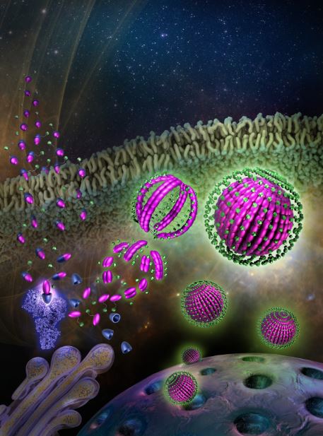
Intracellular self-assembly of OlsaCEST-MRI detectable, DNA methylation-inhibiting theranostic nanoparticles.
The single molecule compound Olsa(green)-CBT (pink)-RVRR (blue) is injected intravenously, passes through the cell membrane with the aid of the cell-penetrating peptide RVRR (representing multiple arginines), and the small peptide substrate RVRR is cleaved by the tumor-associated enzyme furin (purple). The single Olsa-CBT residues are then self-assembled into nanoparticles through a glutathione (GSH) and 2-cyanobenzothiazole (CBT)-mediated biocompatible click condensation reaction followed by π-π stacking.
From: Y. Yuan, J. Zhang, X. Qi, S. Li, G. Liu, X. Song, M.T. McMahon, J.W.M. Bulte. Furin-mediated intracellular self-assembly of olsalazine nanoparticles for enhanced MR imaging and tumor therapy. Nat. Mat. 18, 1376-1383 (2019).
Y. Yuan, P. Raj, J. Zhang, S. Siddhanta, I. Barman, J.W.M. Bulte. Furin-mediated self-assembly of olsalazine nanoparticles for targeted Raman imaging of tumors. Angew. Chem. Int. Ed., doi: 10.1002/anie.202014839.
Magnetizing Cells for Tracking | Science: Out of the Box
Although it has great potential for treating cancer, spinal cord injuries, diabetes and many other serious diseases, there are challenges to making stem cell therapy a reality. Watch cell imaging specialist Jeff Bulte explain his pioneering work in tracking stem cells in the body — and how it can help lead to better stem cell treatments by reporting stem cells’ location and whether they are still alive.

Publications
Pubmed publications Google citations
Neuroscience
The Neuroscience Section is focused on the development of novel MRI technologies and their application to basic science problems and clinical disease, especially in the brain. Methods have and are being developed for MRS and MRS imaging (MRSI); diffusion tensor imaging (DTI) and axonal mapping; physiological imaging (blood flow, blood volume, and blood oxygenation); and for the study of biochemical interactions using magnetization transfer processes.
Other research to improve the understanding of the blood oxygen level-dependent (BOLD) mechanism that underlies functional MRI, and the theory of MR relaxation in blood that underlies it is underway. Several faculty members of this large section are engaged in the development of new technologies for high-field MRI, including novel biodegradable contrast agents (sugar and proteins), molecular imaging markers, and new endogenous contrast agents for distinguishing tumors from healthy tissue.
Our Team
Division Chief
Peter C.M. Van Zijl, Ph.D.

- Chief, Neuroscience, Division of MR Research, Department of Radiology and Radiological Science
- Professor of Radiology and Radiological Science
- Professor of Biomedical Engineering
- Professor of Oncology
-
Susumu Mori, Ph.D.

- Director, Center for Brain Imaging Science, Johns Hopkins Medicine Brain Science Institute
- Professor of Radiology and Radiological Science
- Professor of Oncology


Kazi Akhter, B.S.
Research Associate
[email protected]
Adnan Bibic, Ph.D.
Research Associate
[email protected]
Joe Gillen, B.S.
Research Associate
[email protected]
Wenbo Li, Ph.D.
Assistant Professor
Deng Mao, Ph.D.
Research Associate
Feng Xu, Ph.D.
Research Associate
[email protected]
MRI at Kirby Center
Terri Brawner
MRI Chief Tech at Kirby Center
[email protected]
Kathleen Kahl
MRI Tech
[email protected]
Ivana Kusevic
MRI Tech
[email protected]
Neurofunction Section
Research in the Neurofunction Section is focused on the development of novel MRI techniques to evaluate the brain's physiology, function, metabolism, and structure, as well as their clinical applications. Our work spans across humans to animal models and ranges from neonates to elderly individuals. Our central goal is to develop translatable MRI tools as candidate biomarkers in neurologic and psychiatric diseases.
Representative techniques that our team is actively developing include T2-Relaxation-Under-Spin-Tagging (TRUST) to measure oxygenation and oxidative metabolism, cerebrovascular reactivity (CVR) to evaluate the brain’s vascular reserve, water extraction with phase contrast arterial spin tagging (WEPCAST) for non-contrast assessment of BBB permeability, MR fingerprinting arterial spin labeling (MRF-ASL) for multi-parametric hemodynamic imaging, and brain atlases in special populations such as pediatric patients. Our team is looking forward to opportunities of collaboration and welcomes inquiries or requests of our techniques.
Our Research Lab
Peter van Zijl Laboratory
The Peter van Zijl Laboratory focuses on developing new methodologies for using MRI and magnetic resonance spectroscopy (MRS) to study brain function and physiology. In addition, we are working to understand the basic mechanisms of the MRI signal changes measured during functional MRI (fMRI) tests of the brain. We are also mapping the wiring of the brain (axonal connections between the brains functional regions) and designing new technologies for MRI to follow where cells are migrating and when genes are expressed. A more recent interest is the development of bioorganic biodegradable MRI contrast agents. Our ultimate goal is to transform these technologies into fast methods that are compatible with the time available for multi-modal clinical diagnosis using MRI.
Our Team
-
Hanzhang Lu, Ph.D.

- Elias A. Zerhouni, M.D. Professor
- Director, MRI Service Center
- Chief, NeuroFunction Section, Department of Radiology
- Professor of Radiology and Radiological Science

Neurometabolism
Research in the Neurometabolism Section is focused on the development of novel MRS techniques to measure metabolite levels in the brain, as well as their neuroscience applications. Our work ranges from acquisition to analysis, and from neonates to elderly individuals. Our central goal is to develop tools that make MRS accessible to the non-expert user for applications in clinical and neuroscience applications.
We develop the open-source post-processing and modeling toolkit Osprey, which allows us to advance linear combination modeling of traditional MRS and MRSI data, as well as to develop analyses for the novel acquisition pulse sequence that our group develops. We pioneered the approach of Hadamard-encoded J-difference editing with the HERMES and HERCULES techniques, which allow edited detection of multiple metabolites in a single experiment. We are leading the application of our methods in the HEALthy Baby and Childhood Development (HBCD) study, a national longitudinal study of over 7,000 babies at 25 sites across the US. Our team is looking forward to opportunities of collaboration and welcomes inquiries or requests of our techniques.