In the absence of advanced technology, Wilmer Eye Institute professors relied on the illustrations of dedicated medical artist Annette Smith Burgess to help teach students about diseases of the eye. Combined with a clinical history, Burgess’s beautifully detailed illustrations provided a surprisingly accurate assessment of many eye conditions.
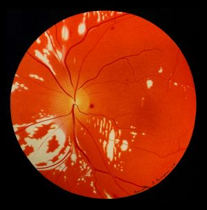
Artfully crafted descriptions further contributed to students’ understanding of anatomical features and could even be poetic in nature: Dr. William Holland Wilmer, describing retinal changes in a diabetic patient in the 1934 Atlas Fundus Occuli (see plate at left), said, “The fine striate edema around the disk has the appearance of a ring around the moon on a misty night."
Today, high-resolution photography allows students to view anatomy and physiology in life-like detail, and residents practice surgical techniques using sophisticated microscopes and 3D simulators in state-of-the-art wet labs. Still, Burgess’s art remains a source of beauty, as well as a reminder of the journey from then to now.