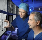
Philippe Gailloud, left, and Rafael Tamargo discuss an arteriovenous malformation. The rare finding requires early intervention for the best outcomes.
Born 5 and a half weeks early, Madalyn Carter was admitted to the NICU at a Washington, D.C. hospital, where neonatologists quickly noticed she wasn’t moving her arms. An MRI revealed the cause—a spinal vascular malformation (SVM) that was compressing the newborn’s spinal cord and cutting off circulation to her brain. At any moment, doctors told her mother, Madalyn was at risk of suffering a severe stroke. Worse, they had no treatment to offer the child.
“We were devastated,” says Meredith Carter. “It was like a ticking time bomb in which she might end up paralyzed.”
Seemingly with nowhere to go, the Fulton, Md., mother remembered that Johns Hopkins pediatric neurosurgeon Ben Carson was a member of her church. She contacted Carson, and he referred Madalyn to pediatric neurosurgeon Rafael Tamargo and interventional neuro-radiologist Philippe Gailloud. Co-directors of the Johns Hopkins Pediatric Stroke and Neurovascular Center, they have deep experience in treating these rare but lethal lesions.
“Experience is important,” says Gailloud. “With it, you have seen them before and you know what you have to deal with.”
“Most doctors, even neurologists and neurosurgeons,” says Tamargo, “will only see a handful of spinal vascular malformations in their entire careers.”
But critical to a good outcomes, say Tamargo and Gailloud, is an early and accurate diagnosis, too. Undetected and untreated, these lesions can grow, bleed and even kill, says Gailloud, noting that dramatically increased pressure in the venous system can cause hypertensive cardiomyopathy.
“When the spinal cord circulation fights against such high venous pressure, everything starts to dilate, and the child gets weak and loses bowel and bladder function,” explains Gailloud. “These very big vascular malformations compress the spinal cord like a tumor, resulting in a bleed or stroke.”
That’s why Tamargo and Gailloud strive to see patients the day after every inquiry they receive on these conditions. But getting patients headed toward the right diagnosis can also be delayed by the difficulty in imaging these conditions. Some spinal fistulas often aren’t visible on MRI, and the ones that show up on CT scan require a shrewd radiologist to spot them.
Definitively diagnosing these problems, Gailloud adds, requires a spinal angiogram—a specialized imaging technique often eschewed by doctors. When this technique was invented in the late 1960s, Gailloud explains, it was blamed for spinal infarctions and a host of other serious complications. Although they still carry a bad reputation, spinal angiograms have become much safer with decades of experience. At Hopkins, Gailloud and his colleagues perform two or three of these procedures each week. They recently published a paper in which they described tracking the complications of spinal angiograms on a series of 350 patients.
“There were no significant complications,” he says. “It turns out to be a very safe procedure.”
Once they diagnose an SVM, Tamargo says, he and Gailloud formulate a treatment plan individualized for each patient. Most can be treated endovascularly, he says, by embolizing their problematic blood vessels with coils or glue. For those whose anatomy or conditions don’t allow endovascular treatment, surgery is the next best option. Madalyn’s case was even trickier. Her SVM—a large perimedullary venous fistula—lied on the interior side of her spinal cord rather than on the back of the neck, making access through the femoral artery the only option. In such cases, says Gailloud, it’s important to stage the procedures.
“To treat this is very tricky—you can damage the spinal cord,” says Gailloud. “If you try to treat the whole thing in one session, the lesion may get so angry that it begins to swell and damage the spinal cord.”
Madalyn underwent her first embolization at 9 months of age, her second at 15 months, both times with no complications. Her outcome?
“Within a day of her first procedure she started to move her arm—it was incredible,” says Meredith Carter. “Now at 4 and a half years old, she’s moving perfectly. You would never know anything was wrong with her. We put her into the right hands.”
“Our goal is to have the lesions recognized earlier so patients have a better chance of a significant recovery,” adds Gailloud. “It’s a crusade for us. The quicker we can address their problems, the better the outcome will be.”
For information or to refer a patient: 410-614-1533.
##