X-rays of the Skull
What is a skull X-ray?
A skull X-ray is an imaging test of the skull bones. X-rays use a small amount of radiation beams to make images. Standard X-rays are done for many reasons. They are done to diagnose tumors, infections, foreign items, or bone injuries.
X-ray beams pass through body tissues onto treated plates. The more solid a structure is, the whiter it looks on the film. Computers and digital media are now more often used in place of films.
X-rays of the skull are not used as often now due to the use of CT scans and MRIs. But they are still helpful for looking for skull fractures and other conditions of the skull and brain.
Bones of the skull
The skull is also called the cranium. It's the bony structure of the head. There are 2 sets of bones make up the skull:
-
Cranial bones. These bones protect and enclose the brain.
-
Facial bones. These bones are the framework for the face and mouth.
All bones in the skull are attached to each other by joints that don't move, except for the jawbone. The jawbone is attached to the skull with a movable joint.
The cranium holds and protects the brain. It's made up of 8 bones. They are:
-
Frontal bone
-
Parietal bones (1 on each side)
-
Temporal bones (1 on each side)
-
Ethmoid bone
-
Sphenoid bone
-
Occipital bone
The face has 14 bones. These include those that make up the jaws, cheeks, and nasal area.
Why might I need a skull X-ray?
X-rays of the skull may be done to diagnose:
-
Fractures of the bones of the skull
-
Birth defects
-
Infection
-
Foreign bodies
-
Certain metabolic and endocrine disorders that cause skull defects
-
Tumors
-
Problems in the nasal sinuses
-
Calcified areas in the brain
There may be other reasons for your healthcare provider to recommend an X-ray of the skull. Be sure to talk with them about the reason for your skull X-ray.
What are the risks of a skull X-ray?
Ask your healthcare provider about the amount of radiation used during the procedure and the risks to you. It's a good idea to keep a record of your radiation exposure to tell your healthcare providers. This includes previous X-rays and CT scans. Risks of radiation exposure may be related to the number of X-ray tests or treatments over time.
If you are pregnant or think you may be, tell your provider. Radiation exposure in pregnancy may lead to birth defects. If you need to have a skull X-ray, care will be taken to protect your baby.
You may have other risks. Ask your healthcare provider before the procedure.
How do I get ready for a skull X-ray?
You don't need to do anything to get ready for a skull X-ray. You don't need to make changes in your food, drink, or medicines. Your healthcare provider will explain the procedure to you and ask if you have questions.
Tell the radiologic technologist:
-
If you are pregnant or think you could be
-
If you have a prosthetic (artificial) eye, because the prosthesis can create a shadow on an X-ray of the skull
What happens during a skull X-ray?
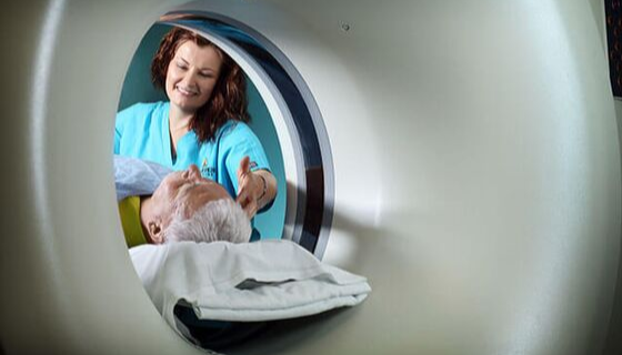
An X-ray may be done on an outpatient basis. This means you go home afterward. Or it may be done as part of your stay in a hospital.
An X-ray of the skull follows this general process:
-
You will be asked to remove any clothing, jewelry, hairpins, eyeglasses, hearing aids, or other metal objects that might interfere with the X-ray.
-
If you are asked to remove clothing, you will be given a hospital gown to wear.
-
You will be positioned on an X-ray table. The technician will make sure that the part of the skull to be X-rayed is between the X-ray machine and a cassette with the X-ray film or a digital plate.
-
A lead apron or shield may be draped over parts of your body that are not to be X-rayed.
-
If the X-ray is being done to find an injury, special care will be taken to prevent more injury. For example, a neck brace may be used if a cervical spine fracture is suspected. The X-ray itself causes no pain. Moving the body into position may cause some discomfort or pain if you have an injury or had surgery. The technician will make sure to minimize any discomfort or pain.
-
The technician will step behind a protective window while the image is taken. They will ask you to hold still for a few moments while the X-ray is taken. The radiation beam will be focused on the area to be X-rayed.
-
Some skull X-ray studies may be done in several different positions. It's very important to be still while the X-ray is taken. Any movement may distort the image and another X-ray may be needed.
What happens after a skull X-ray?
There is no special type of care needed after an X-ray of the skull. Your healthcare provider may give you instructions after the procedure if needed. They will tell you when to expect your X-ray results.
Next steps
Before you agree to the test or the procedure make sure you know:
-
The name of the test or procedure
-
The reason you are having the test or procedure
-
What results to expect and what they mean
-
The risks and benefits of the test or procedure
-
What the possible side effects or complications are
-
When and where you are to have the test or procedure
-
Who will do the test or procedure and what that person’s qualifications are
-
What would happen if you did not have the test or procedure
-
Any alternative tests or procedures to think about
-
When and how you will get the results
-
Who to call after the test or procedure if you have questions or problems
-
How much you will have to pay for the test or procedure