Computed Tomography (CT or CAT) Scan of the Brain
What is a CT scan of the brain?
(Head CT Scan, Intracranial CT Scan)
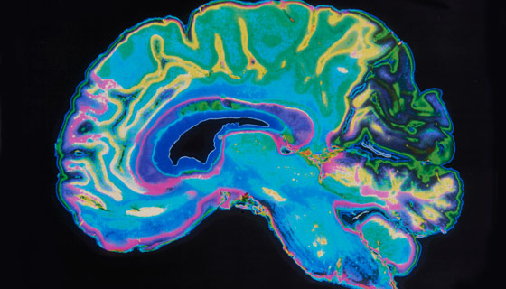
A CT of the brain is a noninvasive diagnostic imaging procedure that uses special X-rays measurements to produce horizontal, or axial, images (often called slices) of the brain. Brain CT scans can provide more detailed information about brain tissue and brain structures than standard X-rays of the head, thus providing more data related to injuries and/or diseases of the brain.
During a brain CT, the X-ray beam moves in a circle around the body, allowing many different views of the brain. The X-ray information is sent to a computer that interprets the X-ray data and displays it in a two-dimensional (2D) form on a monitor.
Brain CT scans may be done with or without "contrast." Contrast refers to a substance taken by mouth or injected into an intravenous (IV) line that causes the particular organ or tissue under study to be seen more clearly. Contrast examinations may require you to fast for a certain period of time before the procedure. Your physician will notify you of this prior to the procedure.
Other related procedures that may be used to diagnose brain disorders include X-rays , magnetic resonance imaging (MRI) of the brain , positron emission tomography (PET) scan of the brain , and cerebral arteriogram .
What is the function of the brain?
As part of the central nervous system (CNS), the brain is an important organ that controls thought, memory, emotion, touch, motor skills, vision, respirations, temperature, hunger and every process that regulates our body.
.png?h=200&iar=0&mh=360&mw=520&w=200&hash=5B9795F96996B95D3B394E0BE48C9744)
What are the different parts of the brain?
The brain can be divided into the cerebrum, brainstem, and cerebellum:
-
Cerebrum. The cerebrum (supratentorial or front of brain) is composed of the right and left hemispheres. Functions of the cerebrum include: initiation of movement, coordination of movement, temperature, touch, vision, hearing, judgment, reasoning, problem solving, emotions, and learning.
-
Brainstem. The brainstem (midline or middle of brain) includes the midbrain, the pons, and the medulla. Functions of this area include: movement of the eyes and mouth, relaying sensory messages (hot, pain, loud, etc.), hunger, respirations, consciousness, cardiac function, body temperature, involuntary muscle movements, sneezing, coughing, vomiting, and swallowing.
-
Cerebellum. The cerebellum (infratentorial or back of brain) is located at the back of the head. Its function is to coordinate voluntary muscle movements and to maintain posture, balance, and equilibrium.
More specifically, other parts of the brain include the following:
-
Pons. A deep part of the brain, located in the brainstem, the pons contains many of the control areas for eye and face movements, facial sensation, hearing, and equilibrium.
-
Medulla. The lowest part of the brainstem, the medulla is the most vital part of the entire brain and contains important control centers for the heart and lungs.
-
Spinal cord. A large bundle of nerve fibers located in the back that extends from the base of the brain to the lower back, the spinal cord carries messages to and from the brain and the rest of the body.
-
Frontal lobe. The largest section of the brain located in the front of the head, the frontal lobe is involved in personality characteristics and movement.
-
Parietal lobe. The middle part of the brain, the parietal lobe helps a person to identify objects and understand spatial relationships (where one's body is compared to objects around the person). The parietal lobe is also involved in interpreting pain and touch in the body.
-
Occipital lobe. The occipital lobe is the back part of the brain that is involved with vision.
-
Temporal lobe. The sides of the brain, these temporal lobes are involved in memory, speech, and sense of smell.
What are the reasons for a CT scan of the brain?
A CT of the brain may be performed to assess the brain for tumors and other lesions, injuries, intracranial bleeding, structural anomalies (e.g., hydrocephalus , infections, brain function or other conditions), particularly when another type of examination (e.g., X-rays or a physical exam) are inconclusive.
A brain CT may also be used to evaluate the effects of treatment on brain tumors and to detect clots in the brain that may be responsible for strokes . Another use of brain CT is to provide guidance for brain surgery or biopsies of brain tissue.
There may be other reasons for your doctor to recommend a CT of the brain.
What are the risks of a CT scan of the brain?
You may want to ask your doctor about the amount of radiation used during the brain CT procedure and the risks related to your particular situation. You should keep a record of your past history of radiation exposure, such as previous CT scans and other types of X-rays, so that you can inform your doctor. Risks associated with radiation exposure may be related to the cumulative number of X-ray examinations and/or treatments over a long period of time.
To safeguard your health, consider the following precautions before scheduling a brain CT:
-
Pregnancy : If you are pregnant or suspect that you may be pregnant, you should notify your doctor. Radiation exposure during pregnancy may lead to birth defects. If it is necessary for you to have a CT of the brain, special precautions will be made to minimize the radiation exposure to the fetus. Contrast media: If contrast media is used during a brain CT, the patient may develop an an allergic reaction to the media. Some patients should not have an iodine-based contrast media. Patients who are allergic to or sensitive to medications should notify their doctor. When you schedule your brain CT scan, you should inform the access center representative if you have had an allergic reaction to any contrast media or if you have kidney failure or other kidney problems. IV contrast will not be administered if you have had a severe or anaphylactic reaction to any contrast media in the past. You may be able to have the scan performed without contrast media or have an alternative imaging exam. . A reported seafood allergy is not considered to be a contraindication for iodinated contrast. Nursing mothers may want to wait 24 hours after contrast material is injected before resuming breastfeeding.
-
Diabetes: Patients taking the diabetes medication metformin (Glucophage) should alert their doctors before having an IV contrast injection as it may cause a rare condition called metabolic acidosis. If you take metformin, you will be asked to stop taking it at the time of the procedure and wait 48 hours after your procedure before restarting this medication. A blood test to check kidney function may be required before you can resume taking metformin.
There may be other risks depending on your specific medical condition. Be sure to discuss any concerns with your doctor prior to the procedure.
How do I prepare for a CT scan of the brain?
If you are having computed tomography angiography (CTA) , you will be given specific instructions when you make your appointment. The following instructions will help you prepare for your brain CT:
-
Clothing : You may be asked to change into a patient gown. If so, a gown will be provided for you. Please remove all piercings and leave all jewelry and valuables at home.
-
Contrast media: You will be asked to sign a consent form that will detail the risks and side-effects associated with contrast media injected through a small tube places in a vein called an intravenous (IV) line. The most common type of brain CT scan with contrast is the double-contrast study that will require you to drink a contrast media before your exam begins in addition to the IV contrast. If you had mild to moderate reactions in the past, you will likely need to take medication prior to the brain CT scan.
-
Food and drink : If your doctor ordered a brain CT scan without contrast, you can eat, drink and take your prescribed medications prior to your exam. If your doctor ordered a CT of the brain with contrast, do not eat anything three hours prior to your brain CT. You are encouraged to drink clear liquids.
-
Diabetics : Diabetics should eat a light breakfast or lunch three hours prior to the scheduled scan. Depending on your oral medication for diabetes, you may be asked to discontinue the use of your medication for 48 hours afterthe brain CT scan. If you have a CT scan with Johns Hopkins radiology, detailed instructions will be given following your examination.
-
Medication : All patients can take their prescribed medications as usual, unless otherwise directed.
Based on your medical condition, your doctor may request other specific steps for brain CT preparation.
.png?h=265&iar=0&mh=360&mw=520&w=175&hash=E75C7F8FA9DD421328CBEB6CD1EE0859)
What happens during a CT scan of the brain?
Brain CT scans may be performed on an outpatient basis or as part of your hospital stay. Procedures may vary depending on your condition and your physician's practices.
Generally, a brain CT scan involves the following steps:
-
If you having a procedure done with contrast, an IV line will be started in the hand or arm for the injection of the contrast media. For oral contrast, you will be given a liquid contrast preparation to swallow.
-
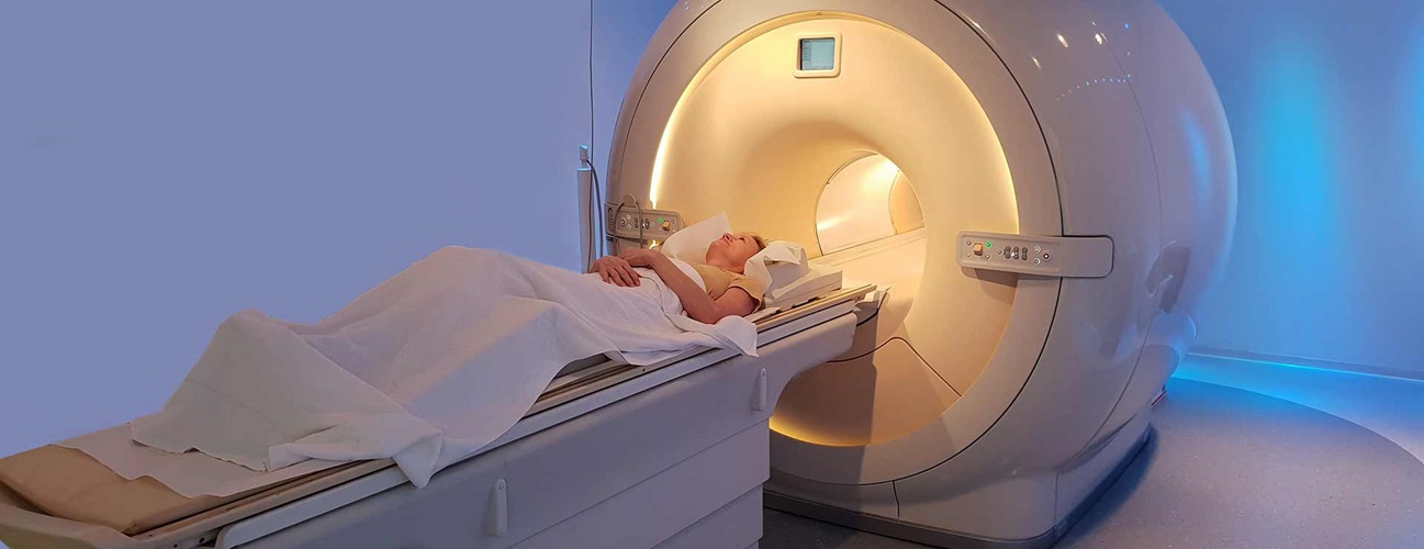
You will lie on a scan table that slides into a large, circular opening of the scanning machine. Pillows and straps may be used to prevent movement during the procedure.
-
The technologist will be in another room where the scanner controls are located. However, you will be in constant sight of the technologist through a window. Speakers inside the scanner will enable two-way communication between the technologist and the patient. You may have a call button so that you can let the technologist know if you have any problems during the procedure. The technologist will be watching you at all times and will be in constant communication.
-
As the scanner begins to rotate around you, X-rays will pass through the body for short amounts of time. You will hear clicking sounds, which are normal.
-
The X-rays absorbed by the body's tissues will be detected by the scanner and transmitted to the computer. The computer will transform the information into an image to be interpreted by the radiologist.
-
You must remain very still during the procedure. You may be asked to hold your breath at various times during the procedure.
-
If contrast media is used for your procedure, you may feel some effects when the media is injected into the IV line. These effects include a flushing sensation, a salty or metallic taste in your mouth, a brief headache or nausea and/or vomiting. These effects usually last for a few moments.
-
You should notify the technologist if you experience any breathing difficulties, sweating, numbness or heart palpitations.
-
When the procedure has been completed, you will be removed from the scanner.
-
If an IV line was inserted for contrast administration, the line will be removed.
While the brain CT itself causes no pain, having to lie still for the length of the procedure might cause some discomfort or pain, particularly in the case of a recent injury or invasive procedure (e.g. surgery). The technologist will use all possible comfort measures and complete the procedure as quickly as possible to minimize any discomfort or pain.
What happens after a CT of the brain?
If contrast media was used during your brain CT scan, you may be monitored for a period of time to check for any side effects or reactions to the contrast media. Notify your radiologist or if you experience itching, swelling, rash or difficulty breathing. If you notice any pain, redness and/or swelling at the IV site after you return home following your procedure, you should notify your doctor as this could indicate an infection or other type of reaction.
Otherwise, there is no special type of care required after a CT of the brain. Most patients are permitted to resume their usual diet and activities. Your doctor may provide additional or alternate instructions after the procedure, depending on your particular situation.