Chordoma
What is chordoma?
Chordomas are tumors that can occur anywhere within the spine or the base of the skull. The two most common locations for chordomas are the lower back (sacral area — approximately one-third to one-half of chordomas) and the base of the skull (approximately one-third of chordomas). Chordomas form from remnants of the notochord — embryonic tissue that eventually forms the center of spinal disks.
These tumors are considered malignant and may metastasize, though they typically grow slowly. Even slow-growing chordomas can become aggressive and grow quite large locally, putting pressure on or invading into critical parts of the brain or spine, which may cause pain and nerve problems or even be life threatening.
What are the symptoms of chordoma?
Chordomas can press on the spine, brain and nerves as they grow, causing pain and nerve problems specific to the part of the brain or spinal cord where they are located. These symptoms can include tingling, numbness, weakness, lack of bladder or bowel control, sexual dysfunction, vision problems, endocrine problems and swallowing difficulties. If the chordoma has grown very large, you may be able to feel a lump.
Chordoma Diagnosis
Treating chordomas can involve extensive surgery, so your doctor will be sure to have a definite diagnosis before planning your individualized treatment.
You will get magnetic resonance imaging (MRI) and computed tomography (CT) scans that will help your doctor determine if any cancer has spread to other parts of the body. You may need to have a needle biopsy, during which your doctor will use a needle to gather a small sample of the tumor for biopsy in order to confirm the diagnosis.
Because the needle biopsy procedure can cause cells of the tumor to spread along the path of the biopsy needle and lead to the cancer spreading, your surgery team will be prepared to remove the tumor and the entire path of the biopsy needle promptly if the pathologist confirms the diagnosis of chordoma.
Chordoma Treatment
When chordomas metastasize (spread) to other parts of the body, or if they grow large and begin to press on critical parts of the brain, they can become life threatening. Even when chordomas have not spread, they can grow very large and damage nerves in the spine and brain, causing disability that may be permanent. It is essential to have chordomas treated promptly while they are still manageable.
Treatment involves preoperative planning, surgery (the procedure is called an en bloc resection, meaning a complete removal of the entire tumor, including any surrounding tissue where cancerous cells may have invaded) and postoperative therapy.
Because of the risk of chordomas spreading, a successful first surgery is very important. Operative intervention provides the best chance for cure and control of the tumor.
Depending on the location, size and appearance of your chordoma, your surgery team might involve multiple surgeons. Chordoma surgery can include a collaboration between specialists in neurosurgery, surgical oncology, orthopaedic oncology, urology, vascular surgery, plastic surgery and anesthesiology. To ensure the best outcome, surgery may also include preoperative planning with medical specialists.
Postoperative care is imperative for the success of your surgery, so after the procedure you will be moved to the neurosurgical intensive care unit (NCCU). In the NCCU, you will be monitored closely to make sure you are recovering well from your procedure. As soon as you are ready, your team will create a plan for physical therapy, occupational therapy, and physical medicine and rehabilitation. After you are discharged from the hospital, you may require a stay in a dedicated inpatient rehabilitation center, where therapists and rehabilitation doctors can continue therapy to maximize your functional recovery.
Your doctor may decide that radiation therapy is necessary to further reduce the tumor’s size or limit its chance of spreading. Radiation treatment involves a collaboration with providers in multiple disciplines, including a radiation oncologist who can help evaluate your case and determine a treatment plan.
Chordoma Ongoing Management
Because of the likelihood of chordoma regrowth, you will need to take charge of your continuing recovery with consistent follow-up.
During the first year after surgery, you will need an MRI every three months to ensure the chordoma isn’t returning. In the years that follow, your doctor may be able to gradually increase the time period between follow-up MRIs.
Kidney Cancer and Chordoma: Craig's Story
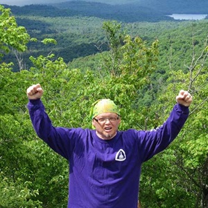
When Craig Scholl was diagnosed with cancer, he and his wife made a trip to The Johns Hopkins Hospital. There, specialists in urology, oncology, neurosurgery and other areas collaborated on a multidisciplinary approach that helped Craig regain his health.