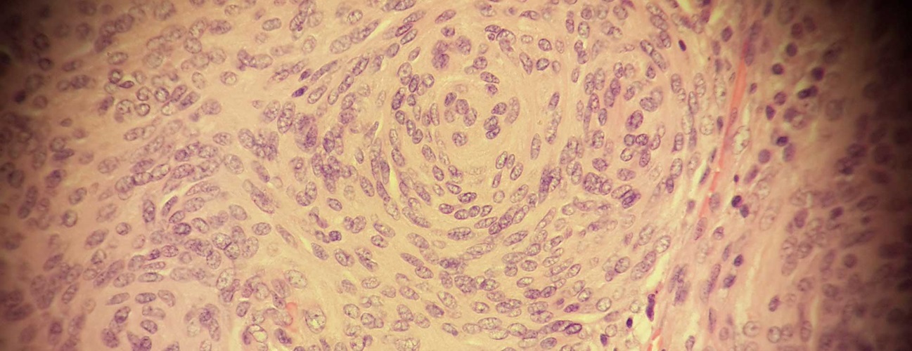
Meningioma cells seen under a microscope

Meningioma

What You Need to Know

  • Meningioma is the most common type of primary brain tumor, accounting for approximately 30 percent of all brain tumors.
  • These tumors originate in the meninges, which are the outer three layers of tissue between the skull and the brain that cover and protect the brain just under the skull.
  • Meningiomas grow out of the middle layer of the meninges called the arachnoid. They grow slowly and may exist for years before being detected. Sometimes doctors will discover a meningioma incidentally on a magnetic resonance imaging (MRI) scan of the head or spinal cord.

Types of Meningioma

Diagram showing where certain meningiomas are located
  • Convexity meningioma grows on the surface of the brain directly under the skull. Accounting for approximately 20 percent of meningiomas, convexity meningiomas may not present symptoms until the tumor has become large enough to push on the brain.

  • Falcine and parasagittal meningioma forms in or next to the falx, a very thin layer of tissue between the two sides of the brain.

  • Intraventricular meningioma forms within the ventricular system in the brain where cerebrospinal fluid (CSF) is made and distributed. An intraventricular meningioma may cause a blockage of CSF flow, leading to hydrocephalus.

  • Skull base meningioma grows in the bones that form the bottom of the skull and in the bony ridge in the back of the eyes. These are more difficult to remove surgically than convexity meningiomas.

  • Sphenoid wing meningioma forms on the skull base behind the eyes. Approximately 20 percent of meningiomas are sphenoid wing.

  • Olfactory groove meningioma forms along the nerves that run between the brain and the nose and account for around 10 percent of meningiomas. This type of tumor can cause a loss of smell, and can grow large enough to cause problems with vision.

  • Posterior fossa / petrous meningioma forms on the underside of the brain and accounts for approximately 10 percent of meningiomas. It can press on the cranial nerves, causing facial and hearing problems. Petrous meningiomas can press on the trigeminal nerve, causing a condition called trigeminal neuralgia.

  • Suprasellar meningioma arises from the base of the skull near the pituitary gland and the optic nerve. Tumors in this area can cause visual problems and dysfunction of the pituitary gland.

  • Recurrent meningioma: Any meningioma may come back. When a meningioma does recur, it may be the same grade or a more aggressive or malignant form.

Find a Doctor

What are the symptoms of meningioma?

If meningioma symptoms occur, they may be very subtle and start slowly as the tumor grows and presses on the brain or spinal cord.

Depending on the meningioma’s size and location, common symptoms may include:

  • Headaches

  • Seizures

  • Blurred vision

  • Weakness in your arms or legs

  • Numbness

Other, less common symptoms include:

  • Loss of balance

  • Hearing loss

  • Memory loss

The Johns Hopkins Brain Tumor Center

Dr. Henry Brem standing in front of an MRI machine.

The Johns Hopkins Comprehensive Brain Tumor Center is one of the largest brain tumor treatment and research centers in the world. We tailor each patient's treatment using an array of advanced approaches, including emerging treatments such as tumor-treating fields and MRI-guided laser ablation.

What are the risk factors of meningioma?

Meningioma is about three times more common in women than in men. The tumors are most common in older patients, with the highest rate in people in their 70s and 80s.

The cause of meningioma is not completely understood, but there are some risk factors:

  • Receiving radiation therapy

  • Having neurofibromatosis type 2, a rare, inherited (genetic) nervous system disorder. People with neurofibromatosis type 2 often get benign tumors of the nerves throughout the body.

Meningioma Diagnosis

  • A physical exam: Your doctor will review your symptoms, personal and family health history, physical exam and tests of vision and reflexes.

  • A neurological exam: This includes assessing your vision, hearing, balance, coordination, reflexes and ability to think and remember.

  • Scans of the brain: MRI and computed tomography (CT or CAT scans) use computers to create detailed images of the brain and are the most common tests used to diagnose brain tumors.

  • Meningiomas have distinct radiological characteristics such as evidence of a dural tail (attachment to the covering of the brain) and indentation of the brain that are likely to show up on these studies.

  • Pathological confirmation: If the tumor is removed, a pathologist will examine the tissue under a microscope to determine the pathological cell characteristics and grade.

Discovering a Meningioma by Accident | Peter’s Story

When Peter fell down stairs and hit his head, he didn’t expect it to save his life. An imaging study revealed a skull base meningioma — a benign tumor of the covering of the brain. He sought care from neurosurgeon Raj Mukherjee, M.D., M.P.H., who performed a minimally invasive craniotomy and removed the tumor.