Lobular Carcinoma in Situ
Featured Expert:
Lobular carcinoma in situ (LCIS) occurs in the linings of milk-producing glands inside the breast — the lobules. Although lobular carcinoma in situ contains the word “carcinoma” (another name for cancer), it is actually not a form of breast cancer. LCIS is a risk factor, however, for developing breast cancer in the future. LCIS is a treatable condition and the outlook is excellent for those who are diagnosed with it.
Breast surgeon Melissa Camp, M.D., M.P.H., explains what you should know about lobular carcinoma in situ and how to work with your doctor to keep yourself safe and healthy.
What You Need to Know
- The phrase “in situ” means “in the original place.” LCIS is contained within the lobules of the breast.
- A typical lobular hyperplasia (ALH), a related condition, is an unusual pattern of cell growth in a localized area of the lobules. ALH, like LCIS, is a marker of increased risk of developing breast cancer in the future. That risk is typically less with ALH than LCIS.
- People with LCIS have approximately a 30% chance of developing breast cancer over the course of their lifetimes, says Camp. The risk depends on their age, life expectancy and other risk factors. (An average woman’s lifetime risk of developing breast cancer, in comparison, is approximately 12%, or about one in eight.)
LCIS Symptoms
Lobular carcinoma in situ does not usually cause symptoms.
“LCIS is typically diagnosed on a breast biopsy performed for an abnormal mammogram, and is only seen on microscopic evaluation of the breast tissue,” Camp says. “There are no specific symptoms or specific mammographic features that correlate with LCIS. It is most commonly an incidental finding.”
Diagnosis of Lobular Carcinoma in Situ
LCIS may be diagnosed with a biopsy. A close look at the LCIS cells by the pathologist can help your doctor decide on the next steps in management.
Breast needle biopsy: “Lobular carcinoma in situ is usually diagnosed with a needle biopsy,” Camp says. “The pathologist [a doctor who examines body tissue for signs of disease] may spot it incidentally when a biopsy is taken for another reason.”
After numbing the breast area, a radiologist or surgeon removes a small amount of breast tissue using a needle. The sample is sent to a pathologist who looks at the tissue under a microscope and can confirm or rule out cancer cells.
“Sometimes, the needle biopsy itself is sufficient,” Camp says. “In some cases, a slightly larger sample of the breast tissue is needed to more definitively rule out breast cancer, and a surgical [excisional] breast biopsy is performed.”
Breast excisional biopsy: “This is a minor procedure performed by a surgeon to remove a slightly larger amount of breast tissue in the location where LCIS was identified,” Camp explains. “It is typically done under general anesthesia or sedation.”
Once you are sedated or asleep, the surgeon makes a cut in the breast and removes part or all of the area of concern. In some cases, the spot may be small, deep and hard to find, requiring the wire localization method. For this, the radiologist or surgeon inserts a needle with a very thin wire into the breast and guides it into the area with LCIS using X-ray images. With the wire in place, the surgeon can identify the area with LCIS and remove a tissue sample for examination under a microscope.
Johns Hopkins Breast Imaging

Johns Hopkins’ breast imaging radiologists are leaders in the field who specialize in breast imaging, reading only breast images. Our state-of-the art breast imaging technology combined with expert interpretation will provide you with the best possible patient care.
Lobular Carcinoma in Situ Treatment
Monitoring and observation may be all that is needed. Camp tells patients diagnosed with LCIS not to panic. “It simply means that we have identified that you may be at higher risk for developing breast cancer in the future,” she says.
“The most important thing to do after diagnosis,” she adds, “is to find a comprehensive breast center where your breast health can be closely monitored and managed.”
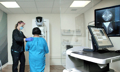
Camp emphasizes the importance of monitoring with imaging so that if breast cancer does develop, it can be detected at the earliest possible stage. “For patients with LCIS, we recommend annual mammograms,” she says, “and we may also suggest annual MRIs of the breast for even closer monitoring.
“In addition, there are medications that can help reduce the risk of developing breast cancer in the future. A discussion with your doctor about these medications can help you decide if they are right for you.”
Breast surgery might be an option if you have other risk factors for breast cancer, such as a BRCA gene mutation or other hereditary factor. Treatment can include a preemptive (prophylactic) mastectomy.
What is the prognosis for lobular carcinoma in situ?
Camp assures people diagnosed with LCIS that the prognosis is very good. “With careful monitoring with an experienced doctor, patients with lobular carcinoma in situ have the best chance to stay healthy and cancer free,” she says.
Johns Hopkins Breast Health Services

Johns Hopkins provides comprehensive world-class care for all conditions related to the breast for optimal breast health. Our services include preventive and noncancerous surgical treatment, risk assessment, diagnostic screenings and treatment for breast cancer.


