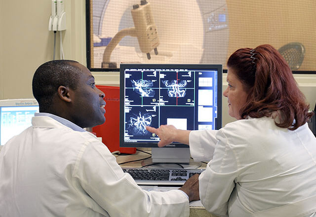
Radiology Exam: Magnetic Resonance Angiography (MRA)
MRA is a diagnostic radiology exam that uses radio waves, a magnetic field and a computer to produce images of inside the body. MRA is used to evaluate blood flow and look for narrowing or blockages of blood vessels to organs or other areas of the body.
To create clearer images of the blood vessels, an MRA exam will often use a contrast agent called gadolinium inserted through an IV line. MRA exams do not use radiation.
MRA: What You Need to Know
- MRA is a type of MRI that looks specifically at blood vessels in the body.
- MRA is often used to examine the blood vessels in the brain, neck, abdomen, heart, chest, arms and legs.
- A doctor may order an MRA exam to look for blood vessel diseases and conditions such as an aneurysm, atherosclerosis, stroke, pulmonary embolism, coronary heart disease, or other abnormalities of the arteries and veins.
- Because an MRA exam produces a strong magnetic environment, it is very important that you tell your doctor about any metal or implantable device anywhere within your body, including a pacemaker, insulin pump or metal pins.
Why Choose Johns Hopkins Radiology for MRA?
Our Physicians
Our diagnostic radiologists have subspecialty training in interpreting MRA images of various areas of the body. Our state-of-the-art equipment and technology are combined with providing the highest level of patient care.
Our Locations
MRA exams may be done on an outpatient basis or as part of a hospital stay. MRA exams are offered at all of our locations. To schedule an exam, call 443-997-7237.
Johns Hopkins Medical Imaging - Green Spring Station
Johns Hopkins Medical Imaging - White Marsh
Johns Hopkins Medical Imaging - Columbia
Johns Hopkins Medical Imaging- Bethesda
The Johns Hopkins Hospital
Johns Hopkins Outpatient Center
Johns Hopkins Bayview Medical Center
