Johns Hopkins is one of only a handful of centers in the world with deep expertise in vertebral column resection (VCR), a complex reconstructive procedure that can correct severe spinal deformities, which often arise as a result of congenital conditions. When multiple vertebral levels are involved, the complexity of the procedure can skyrocket. Indeed, very few physicians worldwide perform VCRs that remove more than two vertebral segments.
Over the span of his career, Johns Hopkins orthopaedic spine surgeon Khaled Kebaish, M.B.B.Ch., has performed roughly two dozen VCRs involving multiple vertebral levels, making him a global expert in the field. Recently, he completed a VCR in which six vertebral segments were removed from a 50-year-old woman with severe spinal kyphosis. Remarkably, there are no published accounts of a VCR of this magnitude in the medical literature.
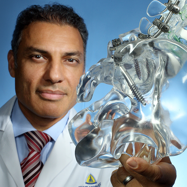
The patient’s spine was nearly folded upon itself, with an abnormal forward curvature of 150 degrees (see photo of 3-D model). Although pain and muscle weakness were not overly prominent, she experienced significant breathing difficulties, requiring her to continually use oxygen, and was unable to eat while sitting. The patient traveled from her home in Kuwait to Johns Hopkins to receive care from Kebaish.
In addition to the procedure’s complexity, the risks to the patient can be significant. They consist of neurologic complications, including permanent spinal cord injury, and life-threatening blood loss. Despite these risks, VCR remains a critical tool for restoring spinal alignment in severe cases—when applied by highly trained surgeons.
In the case of Kebaish’s 50-year-old female patient, he and his surgical team staged her spinal reconstruction in two separate surgeries. The first procedure, lasting roughly five hours, tackled fixation (step one). Following a week or so of recovery, the second procedure, requiring six hours of surgery, was completed, covering steps two through four (see sidebar). The patient fared remarkably well. With VCR, her spinal curvature was restored to near-normal levels (see radiograph on back page).
“Ten years ago, we would not have embarked on a surgery like this,” says Kebaish. “These surgeries, which were previously thought to be impossible, are now made possible by better technologies, planning and understanding of severe spinal deformities through the cumulative knowledge and experience of many people in the field.”
The Role of Referring Physicians
Kebaish stresses the importance of referring physicians as clinical partners. When patients are identified early in the course of their illness as candidates for complex spinal reconstructions, including VCR, their chances of a successful outcome are much higher.
Learn more about the Johns Hopkins Spine Division
at hopkinsmedicine.org/ortho.