Intraoperative CT
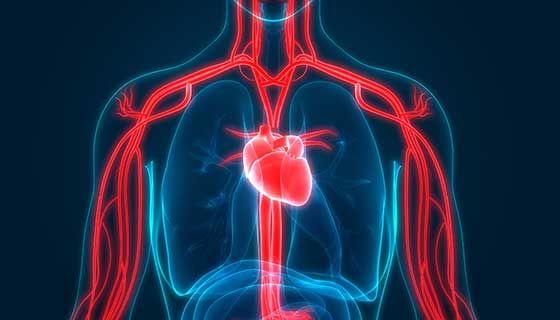
A CT (computerized tomography) scan uses X-rays to produce images of the body. CT scans show bones and organs, as well as detailed anatomy of glands, blood vessels and more.
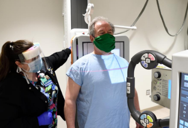
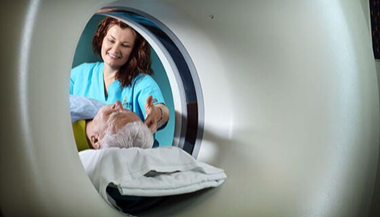
Patients undergo CT scans shortly before surgery to confirm the location of a tumor or other problem and to map the position of vital organs.
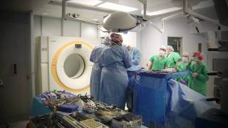
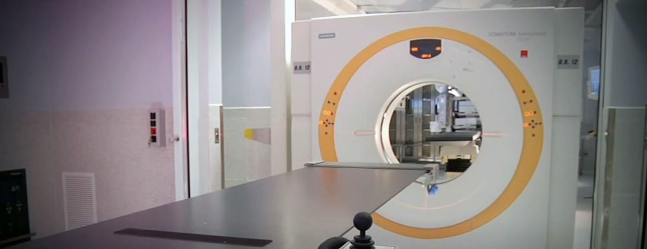
An intraoperative CT (iCT) scanner brings this technology into the operating room, allowing doctors to sync existing scans with new ones. Having access to all of this information at once allows surgeons to better make critical decisions during delicate surgeries, such as those involving the brain.
Benefits of Intraoperative CT
No Need to Leave the Operating Room
The real-time images help verify the success of the operation immediately following surgery, while still in the operating room. Traditionally, the success of a surgery is determined after the procedure is completed and anesthesia has worn off. Verifying during surgery instead reduces the number of procedures patients must undergo. Bringing the CT to patients during surgery eliminates the need to transfer them out of the sterile operating room to a separate location for CT imaging. This lessens the risk of complications that can occur when patients are moved.
Safer Procedures Thanks to Computer Guidance
For neurological surgeries, the iCT is used in conjunction with tools and software that serve as a surgical navigation system. Similar to a GPS device, it allows surgeons to know precisely where they are operating and how much tissue has been removed.
Other Advantages of iCT
- Visualizes changes within the brain as surgery progresses.
- Pinpoints invisible tumor tissue that would otherwise be left behind.
- Identifies dangerous areas of bleeding (hematoma).
- Detects misplaced screws, preventing unnecessary future procedures.
How Intraoperative CT Can Be Used
ICT technology has applications in neurological, orthopaedic and many other types of surgeries, including:
- Procedures that use 3D imaging to determine the precise location of the problem (stereotactic procedures), such as intracerebral hematoma drainage and brain biopsy
- Surgeries involving the face and skull (craniomaxillofacial surgery)
- Hip and pelvic fracture repair
- Internal prosthetics (endoprosthetics) placement, such as joint replacement
- Kidney stone surgery
- Cancerous tumor removal
- Many spine procedures, such as laminectomy, discectomy, vertebroplasty and corpectomy, combined with spinal fusion in the cervical, thoracic or lumbar spine, including:
- Multi-segment fusion for spinal deformity
- Fusion between the top of the spine and the base of the skull (occiput-cervical fusion)
- Fusion between first two vertebrae (atlantoaxial fusion)
- Fusion after disc replacement
- Craniotomy for intracerebral hemorrhage or brain tumor removal
- Insertion of ventriculoperitoneal shunt or intraventricular catheter
- Pituitary tumor removal through the sphenoid sinus
- Subdural hematoma removal