The Departments of Neurology and Neurosurgery provide expert care to thousands of adults and children each year, many with rare, complex conditions. Our team of neurologists and neurosurgeons push the boundaries of what's possible through groundbreaking research and clinical trials.
Schedule an Appointment
Schedule online through MyChart
Schedule by phone
Adult Neurology: 410-955-9441
Pediatric Neurology: 410-955-4259
Adult Neurosurgery: 410-955-6406
Pediatric Neurosurgery: 410-955-7337
International Patients: +1-410-502-7683
Frequently Asked Questions
-
The Johns Hopkins Departments of Neurology and Neurosurgery provide various types of care:
- Specialty care for people who experience issues related to the central and peripheral nervous systems, which include the brain and spinal cord and the nerves throughout the body.
- Inpatient and outpatient care. Outpatient care means that you can see our doctors in a clinic without being admitted to a hospital. Some of our doctors also consult and provide care in the emergency department and various inpatient units throughout our network of hospitals.
- In-person and online care. You can get care in person at one of our locations in the Greater Baltimore or Greater Washington areas. Some clinics may offer online follow-up appointments via telemedicine. If you already use MyChart, you can see if online scheduling is available under specialty care.
-
Our clinicians are experts in a wide range of conditions affecting the nervous system. We treat everything from headaches to rare disorders such as neurofibromatosis. Here are some of our specialty clinics and centers:
-
Treatments for conditions affecting the nervous system vary depending on the diagnosis, medical history and other factors.
Our neurologists specialize in nonsurgical treatments, such as:
- Medications, including infusions
- Lifestyle modifications and symptom management
- Referrals to physical, occupational or speech-language therapy
- Referrals to neuropsychologists and psychologists for cognitive behavioral therapy and similar treatments
Our neurosurgeons specialize in procedures that include open surgical approaches as well as minimally invasive surgeries performed through several small incisions. A procedure may be needed to:
- Remove an abnormal growth, such as a tumor.
- Repair or rewire damaged parts of the brain, nerves or the spinal cord.
- Address bleeding or vascular abnormalities in the brain.
- Install devices in the brain to record and modify brain activity.
- Disable parts of the brain or other neural pathways to stop transmission of abnormal signals.
-
Yes, Johns Hopkins has pediatric neurology and pediatric neurosurgery specialists who treat a variety of nervous system conditions that affect children, including:
- Brain tumors, benign and malignant
- Cerebral palsy
- Epilepsy
- Hydrocephalus
- Spinal muscular atrophy
- Myelitis and myelopathy
- Brachial plexus injuries
- And more
This care is provided as part of Johns Hopkins Children’s Center in Baltimore. Our colleagues at Johns Hopkins All Children’s Hospital in Florida also have dedicated neurology and neurosurgery programs.
-
We have a large team that includes many different medical professionals such as nurse practitioners, physician assistants, neurologists and neurosurgeons. You can find our clinicians in the online directory:
Alternatively, browse our clinics and centers, which list their respective team members.
-
Yes, we offer clinical trials, as research is one of our core pillars at Johns Hopkins Medicine. Some trials are based at Johns Hopkins and are only available here. In other cases, we participate in trials developed by other health institutions.
- Browse open clinical trials for neurological conditions.
- See featured clinical trials in neurology and neurosurgery.
-
You can access Johns Hopkins neurology and neurosurgery services at several locations in the Baltimore metro area as well as Washington, D.C., including:
- The Johns Hopkins Hospital in Baltimore
- Johns Hopkins Bayview Medical Center in Baltimore
- Johns Hopkins Health Care & Surgery Center — Green Spring Station in Lutherville
- Johns Hopkins Health Care & Surgery Center — Bethesda
- Johns Hopkins Community Physicians — Downtown Bethesda
- Sibley Memorial Hospital in Washington, D.C.
Our team performs surgeries at several Johns Hopkins hospitals, including:
-
Johns Hopkins clinicians participate with many commercial health insurance plans.
Learn more about the insurance plans we accept, and be sure to check with your insurer to confirm your plan’s benefits.
Patient Stories
Skull Reconstruction | Dennis' Story

Nerve Repair | Santi’s Story

Surviving Stroke at a Young Age | Sam’s Story


.jpeg)

Hydrogel: The Future of Cancer Treatment
Despite recent technological advancements in the battle against brain cancer, there is a dire need for new treatment strategies. Offering hope in the future of cancer treatment, our team at Johns Hopkins developed a gel to help fend off lingering cancer cells and discourage recurrence after tumor removal.
Read about this research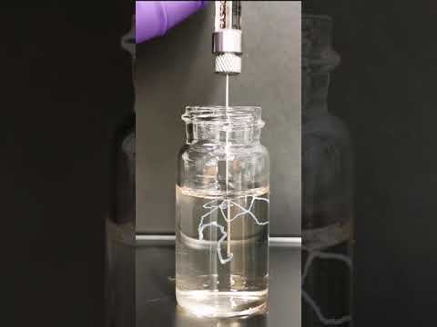
Charitable Giving
-
Neurology
Support future discoveries by Johns Hopkins neurologists.
-
Neurosurgery
Support surgical advancements and discoveries.