Facilities
The Johns Hopkins University School of Medicine provides excellent ambiance for graduate studies. The quality of the research and training activities attracts bright students and outstanding young faculty while maintaining a distinguished senior faculty. The environment, therefore, is very conducive to and supportive of research activities. Almost daily, research seminars are presented by visiting scientists and the latest research development, no matter where it originates, is communicated. Our faculty are editors and reviewers of scientific journals and the aura of serious research is pervasive.
The medical school and hospital environment is especially well suited for research training in human biology. Medical schools are the only schools of human biology and the Johns Hopkins University School of Medicine is among the best places to learn about the human phenotype. The basic science faculty are outstanding human biologists (geneticists, physiologists, pharmacologists, cell biologists and biochemists) and the clinical faculty are experts in human variability and disease.
Transportation
Several shuttle services are provided to connect Hopkins campuses and surrounding areas.
Research Facilities
The research facilities include well-equipped laboratories for molecular biology, cell culture, biochemistry and immunogenetics. Also available are state-of-the-art computer facilities, the Welch Medical Library, with an outstanding collection on genetics; and facilities on the Homewood campus including the Johns Hopkins University Eisenhower Library. The Edward D. Miller Research Building, where the IGM is located, also has a state-of-the-art vivarum with extensive space dedicated to animal research, primarily with rodents.
In addition to laboratory and departmental equipment, several institutional core facilities are available to students. Some of which is available to users after training, and others operate on a fee-for-service basis.
Johns Hopkins Genomics: a premier center for genetics, genomics, and bioinformatics. It is the clinical genetics lab for all campuses of Johns Hopkins Medicine and one of the world’s largest research genotyping and sequencing centers. A partnership between the McKusick-Nathans Department of Genetic Medicine and the Department of Pathology, Johns Hopkins Genomics was established in 2015 to centralize the latest genomic research, the highest quality clinical testing, and exceptional analytic and interpretive expertise. Our mission is to advance the understanding and treatment of human disease, both inherited and acquired, for our patients and colleagues at Johns Hopkins and beyond. Our center is headquartered in Baltimore at 1812 Ashland Avenue in a newly constructed, state of the art laboratory facility. We also have units located in the Johns Hopkins Hospital. Our laboratory units include: the DNA Diagnostic Lab (clinical germline testing), the Molecular Diagnostics Lab (clinical cancer testing), Cytogenetics (clinical testing for germline, cancer, and prenatal chromosome abnormalities), the Genetic Resources Core Facility (research services, consultations, and a CAP-accredited biorepository), and the Center for Inherited Disease Research (high-throughput genotyping and sequencing, statistical genetics, and unrivaled bioinformatics). Our equipment includes the latest NGS, single-molecule sequencing and genotyping platforms. HG Program co-director, Dr. Doheny is the co-director of JH Genomics, opening up rich opportunities for easy access to cutting-edge genomics, bioinformatics, and clinical diagnostic laboratory experiences for our trainees.
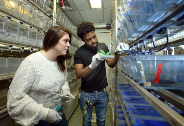
Zebrafish Core Center: Zebrafish are housed in our modern 1500 sq. ft. shared DGM Zebrafish Facility in the MRB. Dr McCallion is the co-director of this facility and its functional genetics (FInZ) CORE. The facility includes room for food preparation and tank changing, as well as bench space with microscopes for sorting embryos and performing procedures such as in vitro fertilization. The facility is outfitted with a 23-rack Aquatic Habitats continuous flow system with continuous water quality monitoring and automated dosing for maintenance of constant pH and salinity. The system has a total capacity for 4000 tanks.
Biostatistics Core: In conjunction with the Biostatistics Center, the Institute for Clinical and Translational Research offers access to a research-related consulting service. Consultants can assist with: research study design, design of data collection systems and instruments, data entry and validation, data management and quality assurance, statistical analysis and data interpretation, and professional and scientific report-writing.
Center for Computational Biology: The Center for Computational Biology (CCB) is a multidisciplinary center dedicated to research on genomics, genetics, DNA sequencing technology, and computational methods for DNA and RNA sequence analysis. The CCB brings together scientists and engineers from many fields, to develop and apply technology that uses sequence data.
CryoEM core: instrumentation and support staff for high-resolution structural studies of proteins and protein and nucleic acid complexes by single particle cryo-electron microscopy and tomography.
JHMI Transcriptomics Core: NextGen sequencing from library construction to data analysis for every major sequencing platform, and for mRNA-seq, ChIP-seq, Single-Read, Paired-End Read, Mate-pair, Targeted Resequencing using Agilent Sure-Select technology, barcode multiplexing. Multiplatform microarray services include Affymetrix, Agilent, Roche/Nimblegen, and Exiqon with associated equipment.
JHMI Synthesis & Sequencing Core: DNA synthesis and purification, automated DNA sequencing, peptide synthesis and purification, and protein/peptide sequencing analysis.
Maryland Advanced Research Computing Center (MARCC): Blue Crab is the main cluster at MARCC with over 23,000 cores (June 2018) and a combined theoretical performance of over 1.4 PFLOPs. The compute nodes combine Ivy Bridge (large memory nodes), Haswell, Broadwell and Skylake processors and several Nvidia K80/P100 GPUS linked via FDR-14 InfiniBand interconnects. It also features two types of storage: 2 PB Lustre (IEEL) and 14 PB ZFS on Linux. The standard compute nodes are Intel Xeon E5-2680v3 (Haswell, 12 cores per CPU), E5-2690v4 (Broadwell, 14 cores per CPU) and Gold 6126 (Skylake, 12 cores per CPU) and 128/96 GB DDR4, 2.5/2.6/2.6 GHz (Marked TDP frequency) or 2.1/2.6/2.3 GHz AVX2(AVX512) base frequency. The large memory nodes are Dell PowerEdge R920 servers with quad Intel “Ivy Bridge” Xeon E7-8857v2, (3.0GHz, 12 core, 30MB, 130W). Each node has 1024 GB RAM.
Mass Spectrometry and Proteomics Core: provides state of the art proteomics services and resources, including: Mass measurements, structural analysis, protein identification, post-translational modification site analysis. Equipment includes QSTAR/Pulsar and LCQ Deca XP Electrospray and Voyager DE-STR MALDI Mass Spectrometers, Nanospray source, Ultimate/Switchos/Famos HPLC, Surveyor HPLC, AP MALDI source for LCQ, IPGPHor and Ettan DALTSix Electrophoresis units, Ettan spot picker, digester and spotter, and Typhoon 9400 Laser Scanner. It provides experimental design advice, courses and training so that interested users may analyze their own samples.
Microscope Facility: Equipment includes 2 transmission electron microscopes (Philips/FEI BioTwinCM120 and Hitachi 7600), a scanning electron microscope (Leo/Zeiss Field Emission), 3 Scanning-spot laser confocal microscopes (Zeiss LSM780-FCS, LSM700, LSM510 Meta), two laser spinning-disk confocal microscopes (3i Marianis/Zeiss Live-cell and 3i/Leica Spinning Disk), a Multiphoton confocal (Zeiss 710NLO Meta), epifluorescence microscopes with stations for TIRF and microinjection, high pressure freezer and freeze substitution system, and general use microscopes for histology and dissection. A laser lightsheet microscope is currently being installed.
Molecular Imaging Center and Small Animal Imaging: PET, SPECT-CT, CT, optical imaging (bioluminescence and fluorescence), MRI and spectroscopic imaging, ultrasound, faxitron (high contrast high magnification), irradiator Kodak multi-spectral imaging, NMR and EPI-MS spectrometry. MRI Service Center: magnetic resonance imaging, spectroscopy and functional imaging. Two 1.5 Telsa imagers with spectroscopy are available, with a 3.0 Tesla imager soon to be added. Multiphoton Imaging Core: Equipped with a wide array of imaging hardware, including: Zeiss LSM 800 microscope with three GaAsP detectors and AiryScan; a Zeiss LSM 880 multiphoton microscope with multiple lasers, AiryScan, time-lapse imaging capabilities, and a near-infrared (IKR) tunable femtosecond Ti-Sapphire laser; a Zeiss 710 multiphoton microscope suitable for awake behaving mouse imaging with multiple lasers, IR imaging capability, a binary GaAsP (BIG) detector and long working distance objectives; a Discovery Laser and Thorlabs Bergamo Multiphoton Microscope; custom, interchangeable temperature-controlled stages; a piezoelectric focus drive for rapid z-focus control; a Zeiss Cell Observer microscope; a Keyence benchtop widefield microscope; a Stereo Zoom Microscope equipped for fluorescent and transmitted light imaging; a stereotaxic surgery station; a 3D printer; and Imaris, Neurolucida, Autoquant, and Zen imaging software.
PhenoCore: pathology support, animal model pathobiology and evaluation, phenotyping strategies, especially for genetically engineered mice.
Protein Microarray Core: full human proteome microarrays and assay design.
Ross Building Flow Cytometry Core Facility offers flow cytometry and high-speed cell sorting to Hopkins researchers.
Single Cell/Nucleus and Data Science Core: The Data Science Core provides end-to-end experimental and computational expertise: initial experimental design and power studies, ongoing data generation and analysis, dimensional data generation and ultimate data deposition and sharing. The Data Science Core will support high and analysis, including multi-omics data integration methods at distinct biological scales.
Synthetic Core: omics data integration method s at distinct biological scales. small molecule synthesis, purification and characterization.
Transgenic Mouse Core: CRISPR/Cas9 mutagenesis, pronuclear injection to produce transgenic mice, blastocyst injection to produce knockout/in m ice, cryopreservation of embryos, rederivitation, aggregation chimeras, derivation of new mouse ES lines, ES cell targeting, and ploidy analysis.
Xray core: states by XProviding the capacity for structural studies of biological macromolecules in crystalline or so ray diffraction and scattering.