-
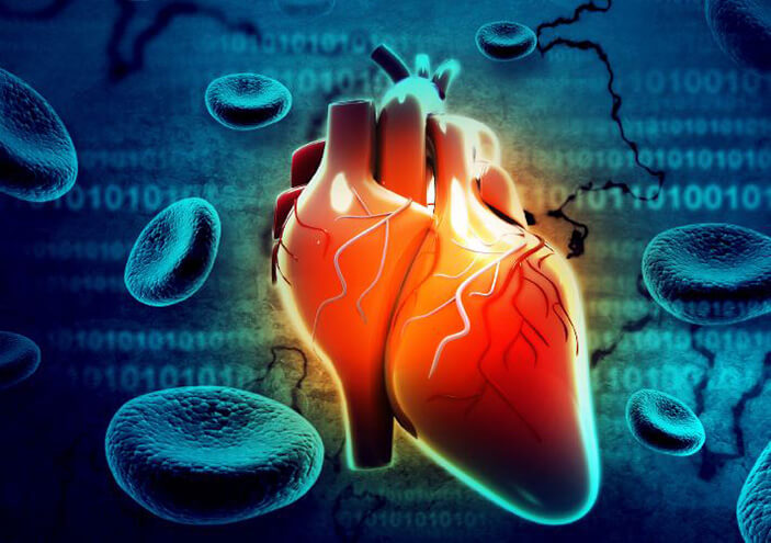
Cardiology
Our team of cardiologists specialize in the health and treatment of the heart.
-
Cardiac Surgery
Johns Hopkins cardiac surgeons perform traditional and minimally invasive heart surgery.
-
Vascular and Endovascular Surgery
Our surgeons treat and care for patients affected by circulatory conditions both common and rare.
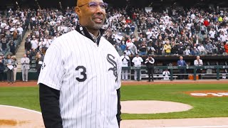
Amyloidosis Leads to Heart and Kidney Transplant: Harold Baines's Story
Harold Baines, MLB Hall of Famer, has a genetic condition called amyloidosis which is a rare disease characterized by a buildup of abnormal amyloid deposits in the body. As a result, he needed both a heart and kidney transplant.

Charitable Giving
-
Cardiology & Cardiac Surgery
Support our advancements and discoveries
-
Vascular Surgery
Support future discoveries by our vascular surgeons