Kidney, Ureter, and Bladder X-ray
What is a kidney, ureter, and bladder X-ray?
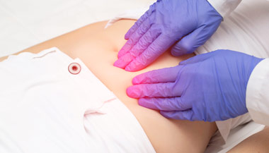
A kidney, ureter, and bladder (KUB) X-ray may be performed to assess the abdominal area for causes of abdominal pain, or to assess the organs and structures of the urinary and/or gastrointestinal (GI) system. A KUB X-ray may be the first diagnostic procedure used to assess the urinary system.
X-rays use invisible electromagnetic energy beams to produce images of internal tissues, bones, and organs on film. X-rays are made by using external radiation to produce images of the body, its organs, and other internal structures for diagnostic purposes. X-rays pass through body tissues onto specially treated plates (similar to camera film) and a "negative" type picture is made (the more solid a structure is, the whiter it appears on the film). Digital films and digital media are more commonly used now than the film media.
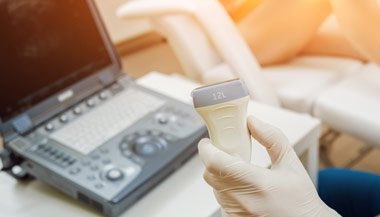
Other related procedures that may be used to diagnose problems of the urinary organs of the abdomen include computed tomography (CT scan) of the kidney , kidney ultrasound , kidney scan , cystography , cystometry , cystoscopy , intravenous pyelogram , kidney biopsy , magnetic resonance imaging (MRI) , prostate ultrasound , retrograde cystography , retrograde pyelogram , uroflowmetry , and renal venogram .
.png?h=132&iar=0&mh=360&mw=520&w=200&hash=852AAE0AC88325AD03DC0A02AFA11A40)
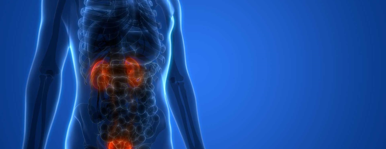
How does the urinary system work?
The body takes nutrients from food and converts them to energy. After the body has taken the food components that it needs, waste products are left behind in the bowel and in the blood.
The urinary system helps the body to eliminate liquid waste in the blood called urea, and keeps chemicals, such as potassium and sodium, and water in balance. Urea is produced when foods containing protein, such as meat, poultry, and certain vegetables, are broken down in the body. Urea is carried in the bloodstream to the kidneys, where it is removed along with water and other wastes in the form of urine.
Urinary system parts and their functions:
-
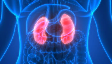
Two kidneys. This pair of purplish-brown organs is located below the ribs toward the middle of the back. Their function is to:
-
remove liquid waste from the blood in the form of urine
-
keep a stable balance of salts and other substances in the blood
-
produce erythropoietin, a hormone that aids the formation of red blood cells
-
regulate blood pressure
-
The kidneys remove urea from the blood through tiny filtering units called nephrons. Each nephron consists of a ball formed of small blood capillaries, called a glomerulus, and a small tube called a renal tubule. Urea, together with water and other waste substances, forms the urine as it passes through the nephrons and down the renal tubules of the kidney.
-
Two ureters. These narrow tubes carry urine from the kidneys to the bladder. Muscles in the ureter walls continually tighten and relax forcing urine downward, away from the kidneys. If urine backs up, or is allowed to stand still, a kidney infection can develop. About every 10 to 15 seconds, small amounts of urine are emptied into the bladder from the ureters.
-
Bladder. This triangle-shaped, hollow organ located in the pelvis. It is held in place by ligaments that are attached to other organs and the pelvic bones. The bladder's walls relax and expand to store urine, and contract and flatten to empty urine through the urethra. The typical healthy adult bladder can store up to two cups of urine for two to five hours.
-
Two sphincter muscles. These circular muscles help keep urine from leaking by closing tightly like a rubber band around the opening of the bladder.
-
Nerves in the bladder. The nerves alert a person when it is time to urinate, or empty the bladder.
-
Urethra. This tube allows urine to pass outside the body. The brain signals the bladder muscles to tighten, which squeezes urine out of the bladder. At the same time, the brain signals the sphincter muscles to relax to let urine exit the bladder through the urethra. When all the signals occur in the correct order, normal urination occurs.
Facts about urine:
-
Adults pass about a quart and a half of urine each day, depending on the fluids and foods consumed.
-
The volume of urine formed at night is about half that formed in the daytime.
-
Normal urine is sterile. It contains fluids, salts and waste products, but it is free of bacteria, viruses, and fungi.
-
The tissues of the bladder are isolated from urine and toxic substances by a coating that discourages bacteria from attaching and growing on the bladder wall.
Reasons for the procedure
A KUB X-ray may be performed to help diagnose the cause of abdominal pain, such as masses, perforations, or obstruction. A KUB X-ray may be taken to evaluate the urinary tract before other diagnostic procedures are performed. Basic information regarding the size, shape, and position of the kidneys, ureters, and bladder may be obtained with a KUB X-ray. The presence of calcifications ( kidney stones ) in the kidneys or ureters may be noted.
There may be other reasons for your doctor to recommend a KUB X-ray.
Risks of the procedure
You may want to ask your doctor about the amount of radiation used during the procedure and the risks related to your particular situation. It is a good idea to keep a record of your past history of radiation exposure, such as previous scans and other types of X-rays, so that you can inform your doctor. Risks associated with radiation exposure may be related to the cumulative number of X-ray examinations and/or treatments over a long period of time.
Notify your health care provider if you are pregnant or suspect that you may be pregnant. Radiation exposure during pregnancy may lead to birth defects.
There may be other risks depending on your specific medical condition. Be sure to discuss any concerns with your physician prior to the procedure.
Certain factors or conditions may interfere with the accuracy of a KUB X-ray. These factors include, but are not limited to, the following:
-
Recent barium X-rays of the abdomen
-
Gas , feces, or foreign body in the intestine
-
Uterine or ovarian masses, such as calcified fibromas of the uterus or ovarian lesions
Before the procedure
-
Your doctor will explain the procedure to you and offer you the opportunity to ask any questions that you might have about the procedure.
-
Generally, no prior preparation, such as fasting or sedation, is required.
-
Notify the radiologic technologist if you are pregnant or suspect you may be pregnant.
-
Notify your doctor and radiologic technologist if you have taken a medication that contains bismuth, such as Pepto-Bismol, in the past four days. Medications that contain bismuth may interfere with testing procedures.
-
Based on your medical condition, your doctor may request other specific preparation.
During the procedure
A KUB X-ray may be performed on an outpatient basis or as part of your stay in a hospital. Procedures may vary depending on your condition and your doctor's practices.
Generally, a KUB X-ray follows this process:
-
You will be asked to remove any clothing, jewelry, or other objects that might interfere with the procedure.
-
If you are asked to remove clothing, you will be given a gown to wear.
-
You will be positioned in a manner that carefully places the part of the abdomen that is to be X-rayed between the X-ray machine and a cassette containing the X-ray film or digital media. You may be asked to stand erect, to lie flat on a table, or to lie on your side on a table, depending on the X-ray view your doctor has requested. You may have X-rays taken from more than one position.
-
Body parts not being imaged may be covered with a lead apron (shield) to avoid exposure to the X-rays.
-
Once you are positioned, the radiologic technologist will ask you to hold still for a few moments while the X-ray exposure is made.
-
It is extremely important to remain completely still while the exposure is made, as any movement may distort the image and even require another X-ray to be done to obtain a clear image of the body part in question.
-
The X-ray beam will be focused on the area to be photographed.
-
The radiologic technologist will step behind a protective window while the image is taken.
While the X-ray procedure itself causes no pain, the manipulation of the body part being examined may cause some discomfort or pain, particularly in the case of a recent injury or invasive procedure, such as surgery. The radiologic technologist will use all possible comfort measures and complete the procedure as quickly as possible to minimize any discomfort or pain.
After the procedure
Generally, there is no special type of care following a KUB X-ray. However, your doctor may give you additional or alternate instructions after the procedure, depending on your particular situation.