Brain Tumor Types
There are more than 120 different types of brain tumors, lesions and cysts, which are differentiated by where they occur and what kinds of cells they are made of. Certain types of tumors are typically benign (noncancerous), while others are typically malignant (cancerous). Others may have a 50/50 chance of being cancerous.
Some of the tumor types listed below may arise from the bone or other types of tissues outside the brain and may also be called “skull base tumors.” However, their proximity to the brain makes them likely to affect structures of the brain, which is why they are included in this list.
- Typically Benign Tumors
- Other Benign Brain Lesions and Cysts
- Brain Tumors with Variable Grades (From More Benign to Malignant)
- Brain Cancer Types: Typically Malignant Brain Tumors
Typically Benign Brain Tumors
Meningioma
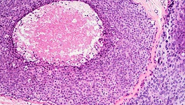
Meningioma is the most common primary brain tumor, accounting for more than 30% of all brain tumors. Meningiomas originate in the meninges, the outer three layers of tissue that cover and protect the brain just under the skull. Women are diagnosed with meningiomas more often than men. About 85% of meningiomas are noncancerous, slow-growing tumors. Almost all meningiomas are considered benign, but some meningiomas can be persistent and come back after treatment.
Pituitary Adenoma
Adenoma, a type of tumor that grows in the gland tissues, is the most common type of pituitary tumor. Pituitary adenomas develop from the pituitary gland and tend to grow at a slow rate. About 10% of primary brain tumors are diagnosed as adenomas. They can cause vision and endocrinological problems. Fortunately for patients affected by them, adenomas are benign and treatable with surgery and/or medication.
Craniopharyngioma
These benign tumors grow near the pituitary gland and can appear as solid tumors or cysts. Craniopharyngiomas often press on nerves, blood vessels or parts of the brain around the pituitary gland. Like adenomas, they can also cause vision and endocrinological issues. They usually affect children and teens as well as adults over the age of 50.
Schwannoma
Acoustic neuromas (vestibular schwannomas) are benign, slow-growing tumors of the nerve that connects the ear to the brain. Less than 8% of primary brain tumors are acoustic neuromas. They usually develop in middle-aged adults, grow on the nerve sheath — the covering surrounding the nerve fibers — and often cause hearing loss. Schwannomas can also affect the trigeminal nerve. These are called trigeminal schwannomas, which are much less common than vestibular schwannomas and can cause facial pain.
Nasopharyngeal Angiofibroma
Nasopharyngeal angiofibroma, also known as juvenile nasopharyngeal angiofibroma, is a benign skull base tumor in the nose that is usually diagnosed in adolescent boys. It is the most common benign tumor of the nasopharynx (the space at the back of the nose that connects the nose with the mouth). It spreads to areas around the nose, causing symptoms such as congestion and nosebleeds.
Choroid Plexus Tumor
Choroid plexus tumors are rare tumors that are found in the choroid plexus — the part of the brain within its ventricles that produces cerebrospinal fluid. About 90% of these tumors are benign. They most frequently occur in children under the age of 2 and can cause hydrocephalus, a buildup of cerebrospinal fluid, as they grow. This can result in increased pressure on the brain and enlargement of the skull. A rare malignant type of choroid plexus tumor is the choroid plexus carcinoma.
Dysembryoplastic Neuroepithelial Tumor
This is a type of neuronal-glial brain tumor — it is made of a mix of neurons and supporting cells. Dysembryoplastic neuroepithelial tumors are rare benign tumors that occur in the tissues covering the brain and spinal cord. Typically found in children and teens, these tumors can cause seizures. Other neuronal-glial brain tumors include gangliogliomas, gangliocytomas and rosette-forming tumors.
Neurofibroma
Neurofibromas are benign, generally painless tumors that can grow on nerves anywhere in the body. In some cases, these soft, fleshy growths develop in the brain, on cranial nerves or on the spinal cord. Multiple neurofibromas are a symptom of a genetic disorder called neurofibromatosis type 1 (NF1).
Hemangioblastoma
Hemangioblastomas are benign tumors of the blood vessels that can form in the brain. These tumors can often be removed through surgery. In rare occasions, they can appear in multiple sites and be symptomatic of a hereditary disease called Von Hippel-Lindau. If that is the case, different tests and visits with a specialist such as an ophthalmologist or geneticist may be recommended.
Chondroma
Chondromas are very rare benign tumors made of cartilage. They can develop in the cartilage found in the skull base and the paranasal sinuses, but they can also affect other body parts such as the hands and feet. Chondromas typically occur in patients between the ages of 10 and 30. While these tumors grow slowly, they may eventually cause the bone to fracture or grow too much, creating pressure on the brain.
Giant Cell Tumor
Named for their extremely large cells, giant cell tumors are rare bone tumors that usually affect the leg and arm bones. They may also be found in the skull. Most giant cell tumors are benign and occur in patients between 20 and 40 years of age.
Osteoma
Osteomas are benign bone tumors (new bone growth) that usually develop on the skull base and facial bones. In general, these slow-growing tumors cause no symptoms. However, if large osteomas grow in certain areas of the brain, they may cause problems with breathing, vision or hearing.
Other Benign Brain Lesions and Cysts
Arachnoid Cyst
Arachnoid cysts are common benign brain cysts that occur in the membranes surrounding the brain and are filled with cerebrospinal fluid. They are usually present since birth, cause no symptoms, and are often left untreated.
Colloid Cyst
This is a benign mass that appears in the third ventricle of the brain. It can block the cerebrospinal fluid pathways, causing headaches and hydrocephalus, or can be found completely incidentally. It is often removed with surgery, especially if it causes hydrocephalus.
Dermoid or Epidermoid Cyst
Dermoid and epidermoid cysts are slow-growing masses that form from leftover skin tissue in embryonal development. They are treated with surgery, and follow-up procedures can be done safely if complete extirpation is not possible during the first surgery.
Encephalocele
Encephalocele is a sac-like protrusion of the brain and the membranes that cover it through an opening in the skull. This rare birth defect occurs when the neural tube, in which the brain and spinal cord form, fails to close completely during fetal development. Encephaloceles can occur in the base of the skull, at the top or back of the skull, or between the forehead and nose. They can cause deformities in the face and cranium that are repaired with surgery. Each year, about one out of every 10,000 infants born in the United States will have encephalocele, according to the Centers for Disease Control and Prevention.
Fibrous Dysplasia
Fibrous dysplasia is a rare bone disorder in which scar-like fibrous tissue develops instead of normal bone. As the bone grows, the fibrous tissue gradually expands, weakening the bone. Fibrous dysplasia usually develops in the skull base and facial bones, thighbone, shinbone, ribs, upper arm bone or pelvis. Fibrous dysplasia can lead to pain and broken or deformed bones. Severe deformity of facial bones can cause loss of vision or hearing. In rare cases, an affected bone area may become cancerous.
Rathke’s Cleft Cyst
Rathke’s cleft cysts are benign fluid-filled growths that slowly develop in the space between the front and back areas of the pituitary gland. Most Rathke’s cleft cysts are found in adults, although they form during fetal development.
Petrous Apex Lesion
Petrous apex lesions are abnormalities that occur in the tip of the bone in the skull next to the middle ear. The most common type of petrous apex lesion is a benign cholesterol granuloma, which is a cyst. Other petrous apex lesions include acoustic neuromas and skull base tumors. Most petrous apex lesions are benign. However, patients with other types of cancer may develop metastatic petrous apex lesions.
Brain Tumors with Variable Grades (From More Benign to Malignant)
Glioma
Glioma is a common type of tumor originating in the brain, but it can sometimes be found in the spinal cord. About 33% of all brain tumors are gliomas. These tumors arise from the glial cells that surround and support neurons. There are several types of glial cells, hence there are many types of gliomas, including:
- Astrocytomas
- A common form is pilocytic astrocytoma, a benign brain tumor arising from the supporting brain cells and usually found in young adults or children. It can be treated with surgery if the entire tumor can be removed.
- Oligodendrogliomas
- Glioblastomas, an especially aggressive tumor type
The understanding of gliomas has been evolving over the years. Depending on the type of cells that are forming the glioma and their genetic mutations, those tumors can be more or less aggressive. A genetic study of the tumor is often performed to better understand how it may behave. For example, diffuse midline gliomas or hemispheric gliomas are newly described types of gliomas that have specific mutations associated with a more aggressive nature.
Ependymal Tumors
Ependymal tumors arise from the lining of the ventricles or the central canal of the spinal cord. They are rare and can appear at any part of the brain or spinal cord. There are different types, including:
- Subependymoma
- Myxopapillary ependymoma
- Ependymoma: Similar to gliomas, ependymomas can also have specific genetic mutations. Their behavior may be different depending on where they are located in the nervous system.
Hemangiopericytoma
Hemangiopericytomas are rare skull base tumors that involve the blood vessels. While most hemangiopericytomas are found in soft tissues of the legs, pelvic area, head and neck, some may occur in the nasal cavity and paranasal sinuses. These tumors may be benign or malignant.
Germ Cell Tumors
During normal development of an embryo and fetus, germ cells usually become eggs (in the female) or sperm (in the male). However, if germ cells travel to the brain by mistake, they can become tumors. Germ cell tumors can be benign or malignant. Some types of germ cell tumors include germinomas, embryonal carcinomas, Yolk sac tumors and teratomas. They are often diagnosed during puberty and tend to affect boys more than girls.
Pineal Tumors
Pineal tumors are those tumors that appear in the region of the pineal gland. They are deep in the brain and can cause hydrocephalus by blocking the cerebrospinal fluid pathways. There are different types of pineal tumors with distinct behaviors, including pineocytomas, pineal parenchymal tumors of intermediate differentiation, and pinealoblastomas. Other tumors such a gliomas, teratomas and germ cell tumors can appear in the pineal region. A blood test or a lumbar puncture may be recommended to look for markers that can help the diagnosis of a pineal tumor. In other instances, a surgery for biopsy is recommended.
Different Types of Brain Tumors

Brain Cancer Types: Typically Malignant Brain Tumors
Chordoma
Chordomas are a rare form of bone cancer usually found in the base of the skull or the lower back. Less than 1% of all primary brain tumors are diagnosed as chordomas. This type of tumor can invade adjacent bone and put pressure on nearby nerve tissue. Although this tumor is slow-growing and looks apparently benign, it behaves more like a malignant tumor because of its tendency to come back and spread to other areas. Chordomas, while generally aggressive, have a somewhat variable and unpredictable progression.
Chondrosarcoma
Chondrosarcoma is a malignant bone cancer that mainly affects cartilage. Chondrosarcoma usually develops in patients between the age of 50 and 70. The tumor can begin in cells in the thighbone, arm, pelvis, knee and spine. It may also start in the face bones or skull base. Some of these cancers can be aggressive.
Medulloblastoma
Medulloblastoma is the most common malignant brain tumor in children. It commonly affects children between ages 5 and 9, and is rare in people over 30. It arises in the cerebellum, a part of the brain located at the base of the skull. There are different types of medulloblastoma, and their aggressivity may change depending on specific mutations of the tumor cells.
Olfactory Neuroblastoma
Olfactory neuroblastoma, also known as esthesioneuroblastoma, is a very rare malignant tumor that develops in the nose. This tumor likely starts in the olfactory nerve, which transmits impulses related to smell from the nose to the brain. Patients with olfactory neuroblastomas may experience frequent nosebleeds, lose their sense of smell and have difficulty breathing through their nostrils.
Lymphoma
Lymphoma is a type of tumor that forms in part of the body’s immune system, the lymphatic system. Lymphomas can spread from other body parts to the brain or form in the brain (primary central nervous system lymphomas). Surgery may be recommended to confirm the tumor type with a biopsy, but primary CNS lymphomas are more often treated with chemotherapy and radiation.
Gliosarcoma
These tumors are rare types of glioma mixed with other supportive tissues. Gliosarcomas can spread to other areas, and are usually aggressive and resistant to therapy.
Rhabdomyosarcoma
Rhabdomyosarcoma is a rare form of soft tissue cancer from the muscle. While it can occur in many places in the body, such as the arms and legs, it is often found in the head and neck and the skull base. Children are more likely to develop rhabdomyosarcomas. This can present with problems in the eyes or face. Certain inherited conditions, including neurofibromatosis type 1 (NF1), increase the risk of this disease.
Paranasal Sinus Cancer
Cancer of the paranasal sinuses forms in the tissues lining the hollow spaces in the bones around the nose. This malignant skull base tumor is primarily found in the maxillary sinuses (in the cheekbones under the eyes) and the ethmoid sinuses (beside the upper nose). There are different types of cancers in the paranasal sinuses and treatment may include a combination of surgery, radiation and chemotherapy.
Atypical Teratoid/Rhabdoid Tumor (AT/RT)
AT/RT is a rare type of embryonal tumor of the central nervous system. It typically affects children 6 and younger, it is fast-growing, and the survival rate is low even with aggressive surgery, radiation and chemotherapy.
The Johns Hopkins Brain Tumor Center
The Johns Hopkins Comprehensive Brain Tumor Center is one of the largest brain tumor treatment and research centers in the world. We tailor each patient's treatment using an array of advanced approaches, including emerging treatments such as tumor-treating fields and MRI-guided laser ablation.