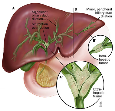
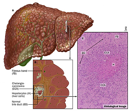
Bile Duct Cancer (Cholangiocarcinoma)
Cholangiocarcinoma, or bile duct cancer, occurs when a malignant (cancerous) tumor grows in one of the ducts that transport bile from the liver to the small intestine.
Bile Duct Cancer Symptoms
Symptoms depend on the location of the tumor or tumors. Symptoms can include:
-
Jaundice (yellowing of the skin and whites of the eyes)
-
Pruritus (itching)
-
Abdominal pain
-
Fever
-
Weight loss and/or progressive weakness
-
Distended gallbladder
Bile Duct Cancer Diagnosis
A diagnosis of bile duct cancer begins with comprehensive physical exam during which you describe your symptoms and medical history. Other tests your doctor may perform include:
-
Laboratory Tests
-
Imaging Scans
-
Endoscopic Diagnosis
-
Percutaneous Radiological Diagnosis
Laboratory Tests
Blood tests will check your liver function and look for markers that may indicate a tumor. Tests include:
-
Liver function tests
-
CEA and CA19-9, blood tests that check for underlying gastrointestinal malignancies
-
Alpha-Fetoprotein (AFP), a blood test that is used to identify a possible malignancy. Used to diagnose liver cancer, it is also helpful in diagnosing bile duct cancer.
Imaging Scans
Imaging scans are noninvasive, painless procedures that your doctor uses to obtain detailed images of the inside of your body. Each type of scan utilizes a different kind of technology to obtain the images.
Scans include:
-
Transabdominal ultrasound. This is usually the first test you will undergo if bile duct cancer is suspected. During an ultrasound, sound waves bounce off your internal organs and tissue to create an image.
-
Computed tomography (CT) scan. A CT scan is a very powerful X-ray and can detect masses associated with bile duct cancer.
-
Magnetic resonance imaging (MRI) . An MRI is slightly superior to a CT scan for visualizing bile duct tumors. An MRI uses magnetic waves to create a detailed image. The latest MRI technology, magnetic resonance cholangiography (MRCP), is a specific type of MRI used to help diagnose bile duct cancer.
-
Positron emission tomography (PET) scan. A PET scan is a nuclear medicine imaging technique. Your doctor will inject you with a low dose of radioactive sugar that concentrates around abnormalities such as tumors. PET scans can be used to detect bile duct cancer. A PET scan and CT scan may be performed at the same time.
Endoscopic Diagnosis
An endoscope is a thin, flexible, lighted tube used to view your upper gastrointestinal tract. Your upper GI tract includes the esophagus, stomach and duodenum (the first part of your small intestine). An enteroscope, a specialized endoscope, allows your doctor to view an even larger part of your small intestine.
There are a number of endoscopic procedures, including:
-
Gastrointestinal endoscopy . You receive a topical anesthetic, as well as pain medication and a sedative if necessary. Your doctor inserts the endoscope through your mouth and into your esophagus. The endoscope is equipped with a camera, which sends images to a monitor for viewing.
-
Endoscopic retrograde cholangiopancreatography (ERCP) . An ERCP uses a special side-viewing endoscope. This endoscope allows your doctor to inject contrast material into your bile ducts. X-ray pictures are taken and the contrast highlights any abnormalities. During an ERCP, your doctor can perform a biopsy and remove bile duct tissue for analysis.
-
Cholangioscopy . A cholangioscope is a special type of endoscope that your doctor inserts inside the bile duct. The cholangioscope is used to see and biopsy the tumor directly. You may have a cholangioscopy done during an ERCP.
-
Endoscopic ultrasound (EUS) . This procedure combines an endoscopy with an ultrasound to obtain images from your gastrointestinal tract. The procedure is similar to an upper endoscopy. EUS is useful in diagnosing bile duct cancer.
Percutaneous Radiological Diagnosis
Percutaneous radiological diagnosis is a more invasive type of imaging scan. Percutaneous refers to something penetrating the skin. A radiologist obtains images after inserting a needle into the affected area.
There are two types of percutaneous radiological diagnosis:
-
Percutaneous transhepatic cholangiography . Your doctor inserts a small needle through your liver and into one of the biliary ducts. He or she injects contrast material through the needle, and then X-ray images are taken in order to see the biliary tree. In some cases, a percutaneous transhepatic cholangiography is more useful than an ERCP.
-
Angiography. This procedure allows your doctor to stage a tumor prior to surgery. Using this procedure allows he or she to evaluate the severity of the biliary obstruction and determine resectability, which means the ability to remove the abnormality through surgery. It also shows all the veins and arteries that will be affected by the surgery.